
<p>This man was referred as a 2-week wait with a skin lesion on the right upper back. What is the diagnosis?</p>
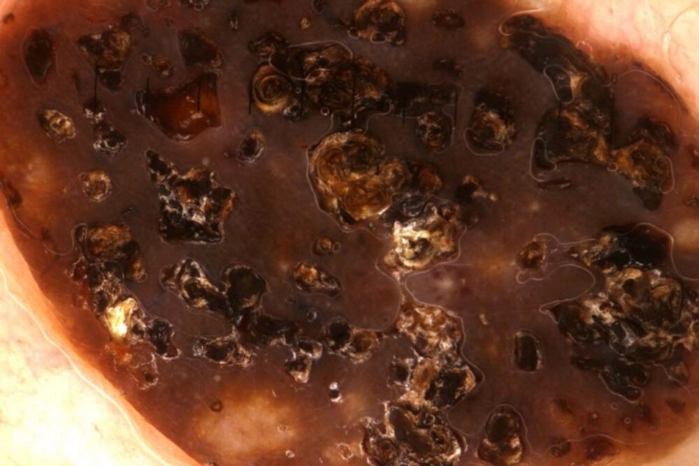
<p>There are many large grainy clods in keeping with a seborrhoeic keratosis. There are no dermoscopic features of a melanocytic lesion.</p>
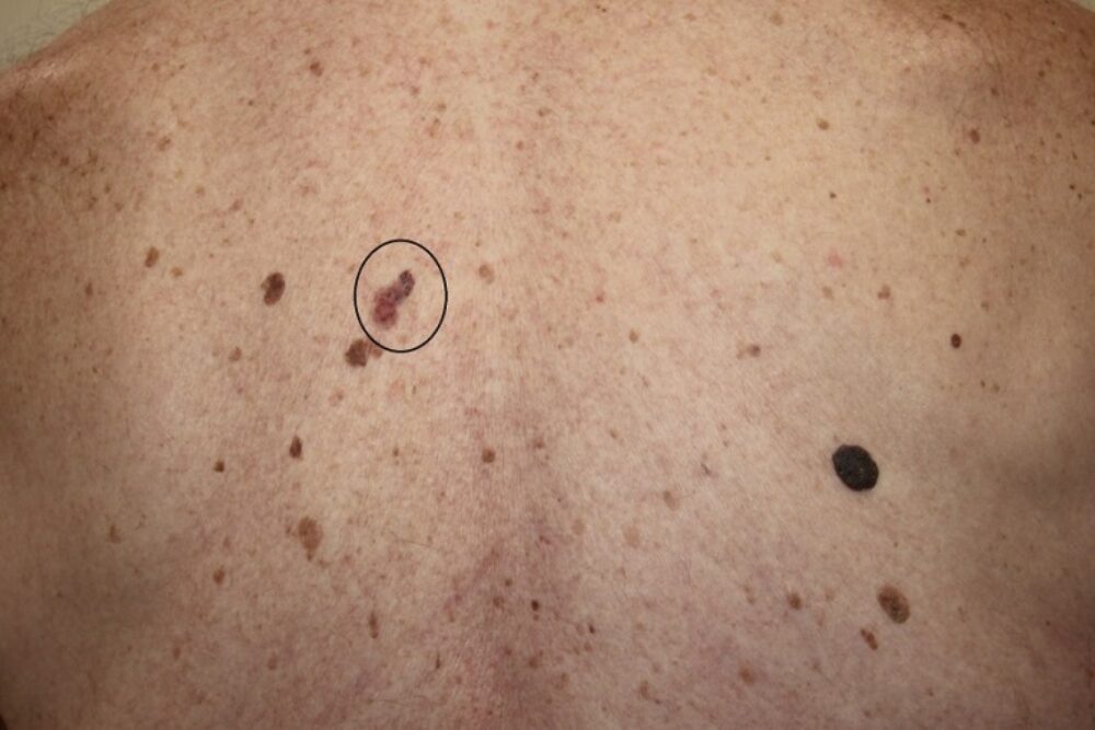
<p>Further examination revealed the real ugly duckling, which is on the left upper back.</p>

<p>What is the diagnosis?</p>
<p>There are no obvious features of a melanocytic lesion (ie pigment network, <strong>aggregated brown/black dots and rounded clods</strong>, peripheral lines), but there are blue clods (black circle) and many <strong>large peripheral irregular brown clods</strong> (white circle) in keeping with a BCC. In general, the irregular brown clods in BCC tend to be seen at the periphery (and again there is nothing that I could call a leaf-like structure if we were using a metaphorical dictionary). Given that we see blue in this BCC we know it cannot be superficial and so surgery is required.</p>
<p>So often we find a patient referred in with one lesion, who goes out with a different lesion in the specimen pot. </p>
