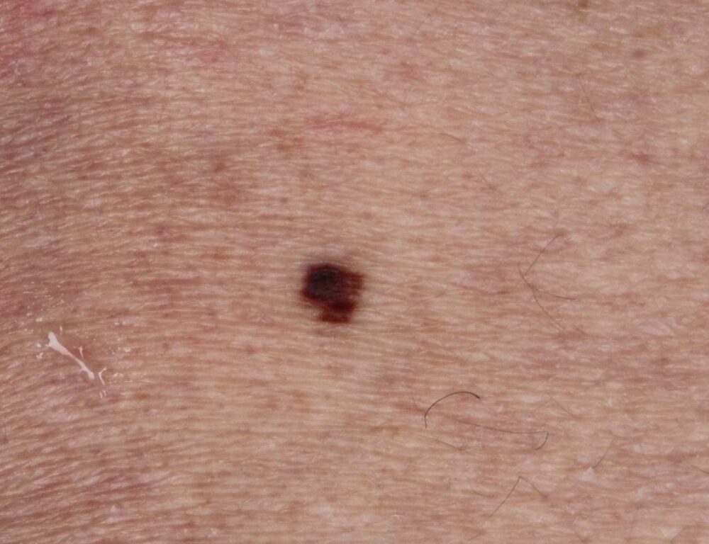
<p>A 68 year old man was noted by chance to have a pigmented lesion on his left upper back, he was uncertain how long it had been present for and whether it had changed. </p>
Dermoscopy - Introduction, Terminology and Structures
A document to help you understand the dermoscopic descriptions used in this blog.
Dermoscopy Algorithm
To help you work through dermoscopic cases.
<p>Given the dermoscopic features how are you going to manage this patient?</p>
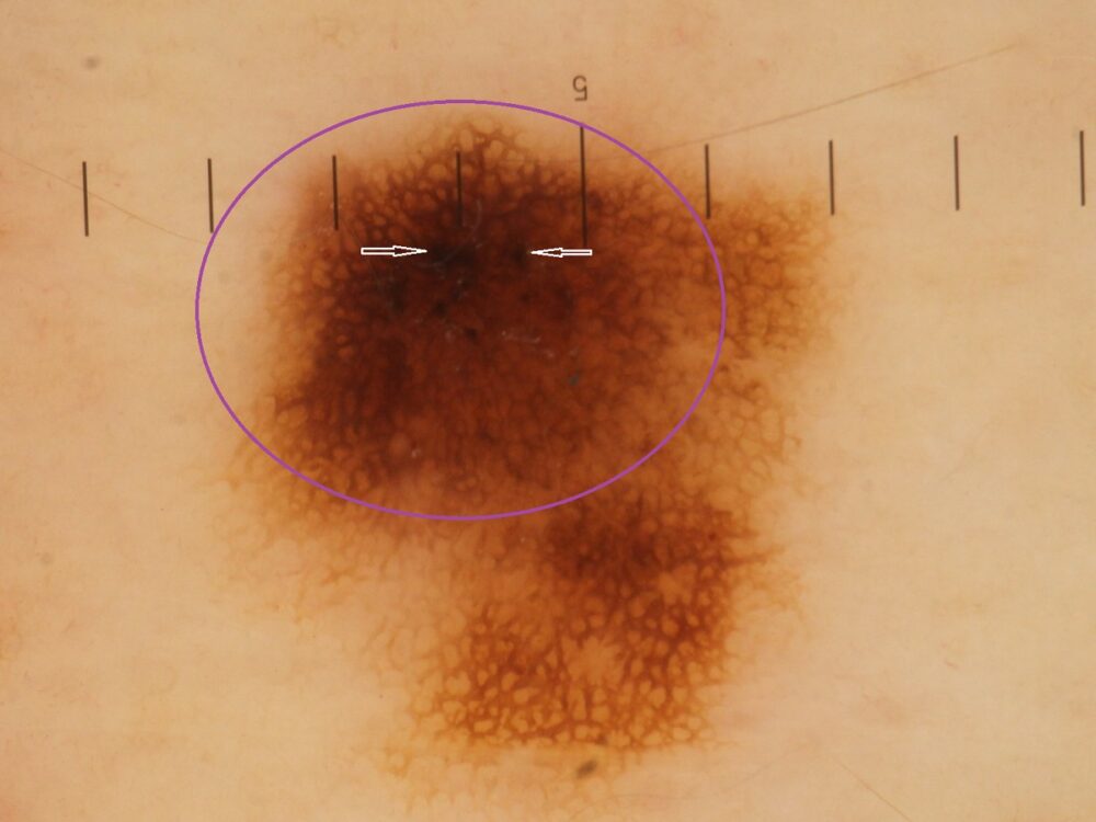
<p>The lesion is clearly asymmetrical and looking at the circled area the network is thicker and darker compared to the rest of the network suggesting that this component is atypical. The lesion was excised and there was found to be an area of severe dysplasia (purple circle) on a background of mild dysplasia. The excision margin of 5 mm was sufficient.</p>
<p>Most pigment networks are clearly typical, while a few are obviously atypical, however, in some cases it is not clear, after all such assessments are subjective. To try and help decide whether a network is atypical, and whether or not the lesion needs to be excised, it is worth considering a number of factors including:</p>
<ul><li><strong>Asymmetry of dermoscopic features</strong> - the greater the degree of asymmetry the greater the level of concern</li><li>Are there <strong>other atypical features? </strong>in this case the blacks clods (white arrows) were atypical </li><li>Is the lesion new or old and changing vs long-term unchanged - in this case there was no clear<strong> history</strong> as this was on the back</li><li><strong>Age</strong> - the risk of skin cancer increases with age</li><li><strong>Pattern comparison </strong>- is perhaps the most important feature, are there other naevi that have have the same dermoscopic pattern, or as with this lesion is it an ugly duckling?</li></ul>
<p>Of course something else that helps greatly is experience - the more you practice dermoscopy the better you will become. </p>
<p>Images copyright of South Tees Hospitals NHS Foundation Trust. </p>
