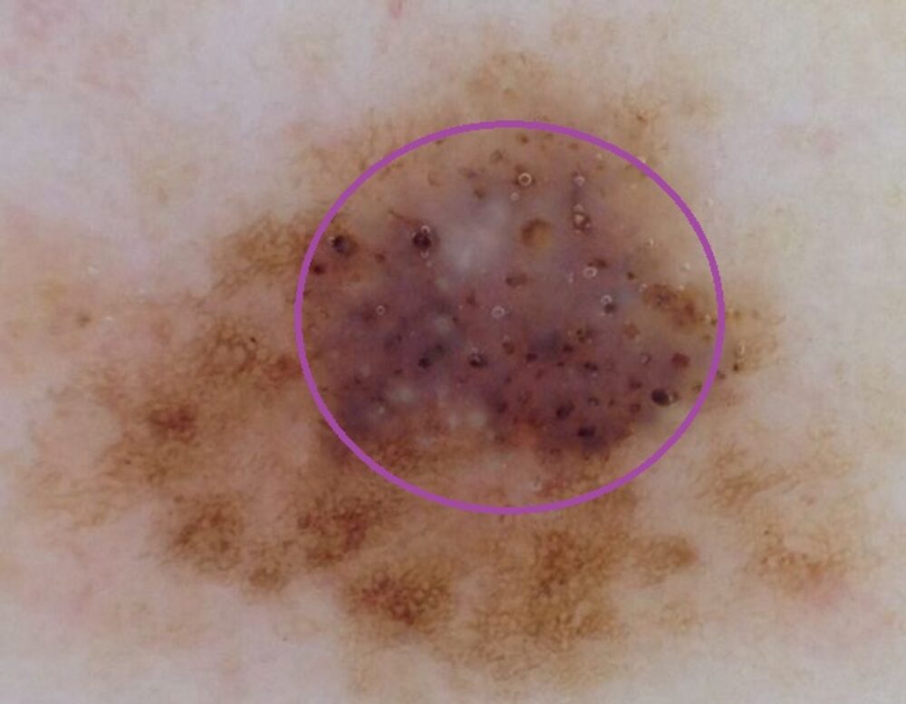
<p>There is a pigmented network so we know the lesion is melanocytic. In addition there are many white clods and grainy brown clods (circled), but why? Essentially this is what is referred to as a collision tumour, where there are two different lesions seen together. In this case the patient had a long-term melanocytic lesion, on top of which grew a seborrhoeic keratosis. </p>
