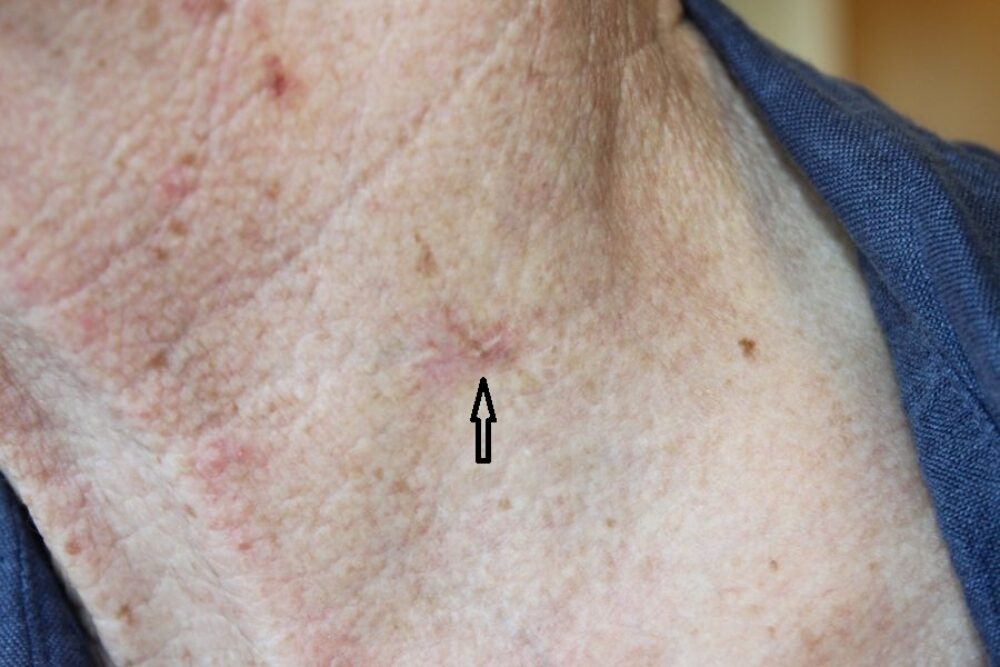
<p>This lady was noted by chance to have a subtle skin change to the left side of her neck. Clinically there is not a lot to go on, but dermoscopy reveals all we need to know. </p>

<p>Dermoscopically we see many fine, well-focused, branching vessels in keeping with a BCC. The lesion felt very flat and so the question was did this need to be excised or could we offer non-surgical treatments?</p>

<p>As we move the dermatoscope along the lesion we start to see blue-grey clods, an important clue in management terms because <strong>BCC with blue-grey clods are less likely to respond to non-surgical approaches. </strong>In terms of our patient it was therefore decided that the lesion should be excised with a 4 mm margin. </p>
<p>An article in Clinical and Experimental Dermatology (2018) formulated that:</p>
<ul><li><strong>Superficial BCC </strong>can be seen dermoscopically as having irregular peripheral brown clods (maple leaf-like structures) and short fine vessels, which in my experience tend to be linear and arranged more at the periphery<br></li><li><strong>Non-superficial BCC</strong> can be seen dermoscopically as having blue-grey clods and arborising vessels </li></ul>
<p>Images copied with kind permission from South Tees Foundation Trust. </p>
