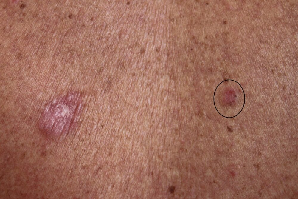
<p>A 69 year old man was referred to clinic. He had very UV-damaged skin and a thorough skin examination highlighted many relevant lesions. Two of these lesions, both of which were on his upper back, were the same diagnosis. I have circled the lesion on the right for further discussion.</p>

Dermoscopy - Introduction, Terminology and Structures
A document to help you understand the dermoscopic descriptions used in this blog.
Dermoscopy Algorithm
To help you work through dermoscopic cases.
<p>Dermoscopcially much of the lesion is pink and structureless. There were a number of erosions (white arrows) that had crusted over, and a few grey structures at the periphery (white circles), which appeared more grey in clinic than compared to the image shown here. <strong>What is the diagnosis?</strong></p>
<p>All the features taken together are in keeping with a superficial BCC. Indeed the patient was found to have some 10 other sBCC, 3 solid BCC and a melanoma with Breslow thickness of 0.9 mm on the left forearm. </p>
<p>With so many BCC<strong> how should we manage such a patient?</strong> The first thing is to be confident about the diagnosis, any lesions that are less than typical require a biopsy. Once satisfied then options need to be discussed with the patient - clearly surgery is unlikely to be practical or in the patient's best interests for so many lesions, except the 3 solid lesions. Choices for the sBCC include topical treatments (Efudix or imiquimod), photodynamic therapy, or even watch and wait and only treat those that become symptomatic.</p>
