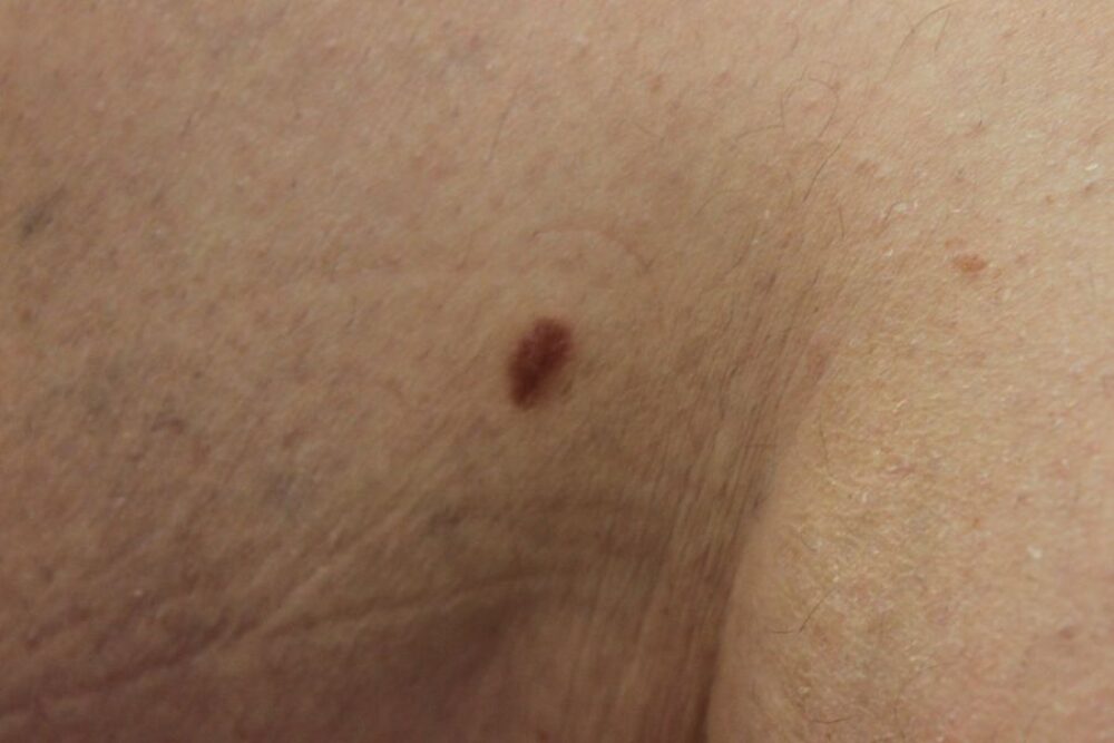
<p>A 56 year old lady presented with a changing pigmented lesion on the left lower inner thigh. What is the differential diagnosis?</p>
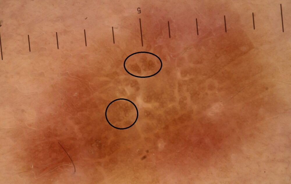
<p>Clinically the lesion could be melanocytic (benign or malignant) or a dermatofibroma.</p>
<p>Dermoscopically there is a <strong>central symmetrical white network</strong> and a <strong>subtle brown rounded network (black circles)</strong>, all in keeping with a dermatofibroma. </p>
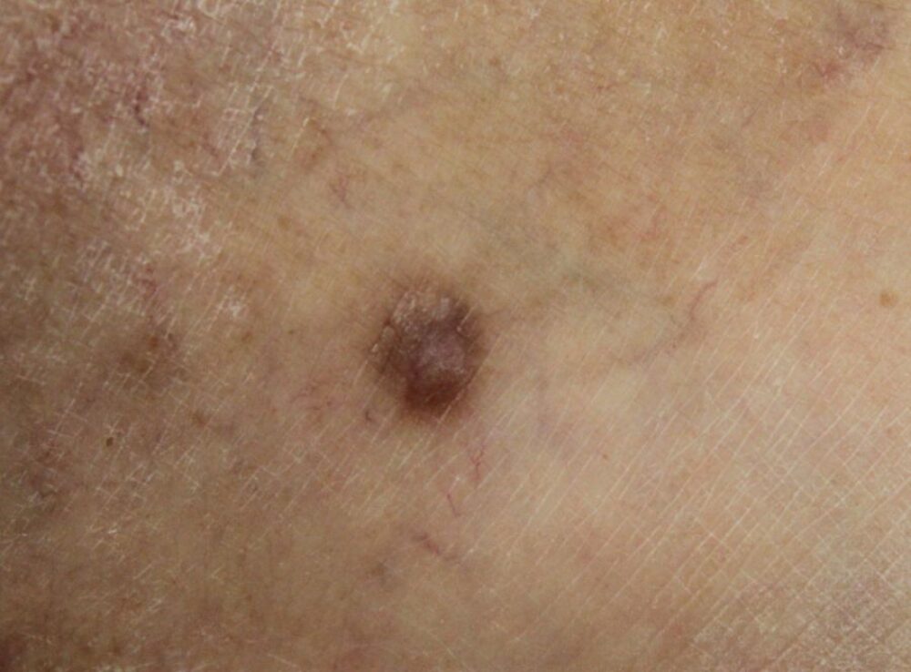
<p>On the ankle of the same leg was a very similar looking lesion.</p>
Dermoscopy - Introduction, Terminology and Structures
A document to help you understand the dermoscopic descriptions used in this blog.
Dermoscopy Algorithm
To help you work through dermoscopic cases.
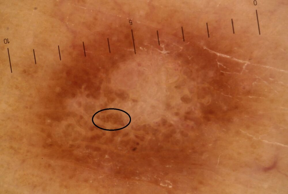
<p>On this occasion we see a central white structureless area, a rounded brown network is present but you need to look more carefully. Again another dermatofibroma.</p>
<p><strong>Dernatofibromas are generally consistent in their behaviour and appearance:</strong></p>
<p>- Grow slowly to reach a diameter usually of less than 1 cm<br>- Limbs most common site<br>- Can be solitary or several <br>- Brown-red in colour, sometimes with a slightly more pronounced brown at the periphery<br>- Often palpable, with a firm symmetrical deeper lump under the surface<br>- Dermoscopically a white centre that can be structureless, a network, or short white lines. Areas of a fine brown rounded network, most commonly at the periphery. Flatter lesions have little in the way of a white centre, and more in the way of a network</p>
<p>Images copyright of South Tees Foundation Trust.</p>
