A 17 year old lady was referred with a new lesion on her thigh. There was no other relevant personal or family history. How would you manage the patient and what is the differential?
Dermoscopy - Introduction, Terminology and Structures
A document to help you understand the dermoscopic descriptions used in this blog.
Dermoscopy Algorithm
To help you work through dermoscopic cases.
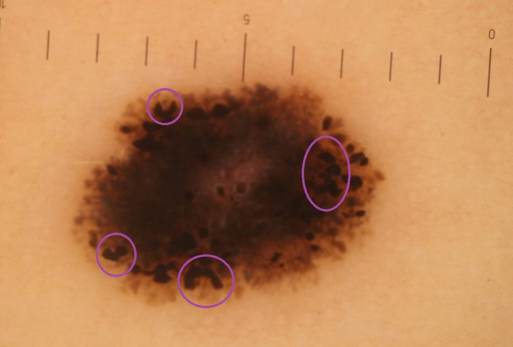
This lesion certainly required a 2-week wait referral, given the clinical and asymmetrical dermoscopic pattern urgent histology was clearly required.
The young age of the patient along with layered peripheral clods points to a pigmented Spitz naevus. There are many C shaped clods, a feature I have seen in some Spitz naevi. Given the significant variation in shape, size and distribution of clods then an atypical spitz / melanoma could not be excluded on clinical grounds alone.
The diagnosis was a pigmented Spitz naevus.
The term Spitz naevus was first introduced in 1948 by Sophie Spitz, at that time such lesions were considered melanomas of childhood.
Images copied with kind permission from South Tees Hospitals NHS Foundation Trust.