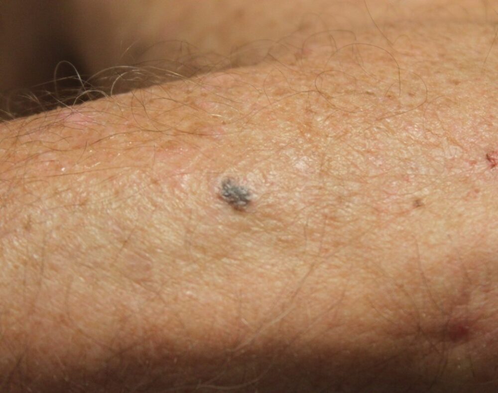
A 67 year old man was referred with a skin lesion that turned out to be nothing particularly untoward. However, there was very noticeable UV damage so I went on to perform a full skin examination, which revealed this lesion on his left forearm that he was uncertain of in terms of history.
Dermoscopy - Introduction, Terminology and Structures
A document to help you understand the dermoscopic descriptions used in this blog.
Dermoscopy Algorithm
To help you work through dermoscopic cases.
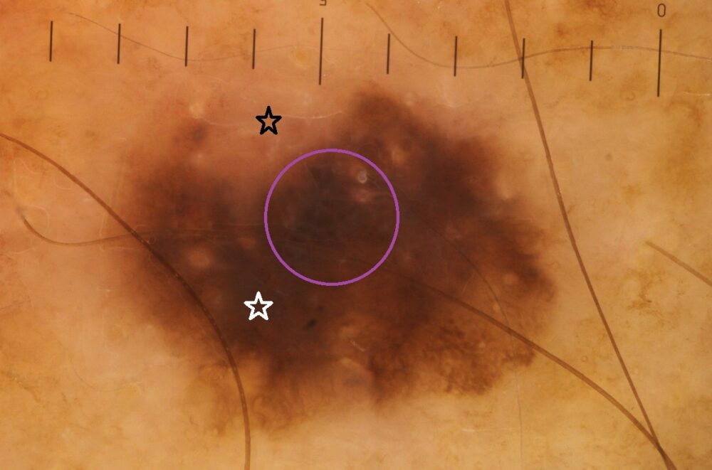
Dermoscopically there is the pigment network (lower-right pole) of a melanocytic lesion. There is asymmetry with eccentric structureless areas (black star = white area, white star = brown area), which as you can see from the attached algorithm is cause for concern. Also, if you look closely there is an atypical white network (purple circle).
The lesion was excised and the diagnosis a superficial spreading melanoma with a Breslow thickness of 0.9mm.
While in general practice teledermoscopy has a potential role for solitary lesions it does not come close to the gold standard set by those specialised in clinicodermoscopic examination undertaking a thorough skin assessment, especially in those cases with background actinic damage and/or large numbers of lesions. A teledermoscopic referral of the initial lesion on the back may have meant that this melanoma on the forearm would not have been picked up as early as it was.
Copied with kind permission from South Tees Foundation Trust.