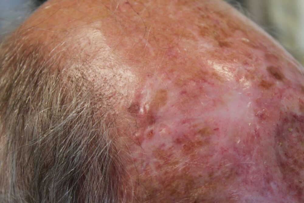
<p>This patient from South Africa is under the care of both dermatology and plastic surgery. He has had multiple skin cancers excised from the scalp. Can you see where the problem is today?</p>
Dermoscopy - Introduction, Terminology and Structures
A document to help you understand the dermoscopic descriptions used in this blog.
Dermoscopy Algorithm
To help you work through dermoscopic cases.
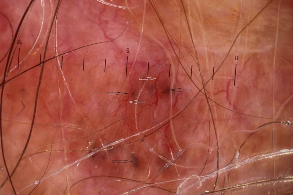
<p>Dermoscopically there are <strong>well-focused branching (arborising) vessels</strong> (white arrows) and <strong>blue-grey clods</strong> (black arrows) in keeping with a <strong>BCC.</strong></p>
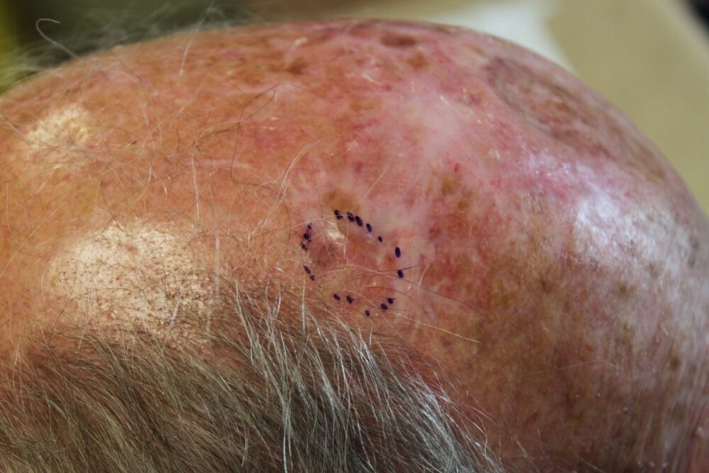
<p>Clinically the lesion was not obvious, but the dermatoscope came to the rescue. With all the previous surgery the skin was very tight so the lesion could not be excised by primary closure. I referred the patient to my plastic surgical colleagues and uploaded an<strong> image of the dermoscopic limits of the BCC</strong> on to the hospital image storage system to aid the plastic surgeons.</p><p>Copied with kind permission from South Tees NHS Foundation Trust. </p>
