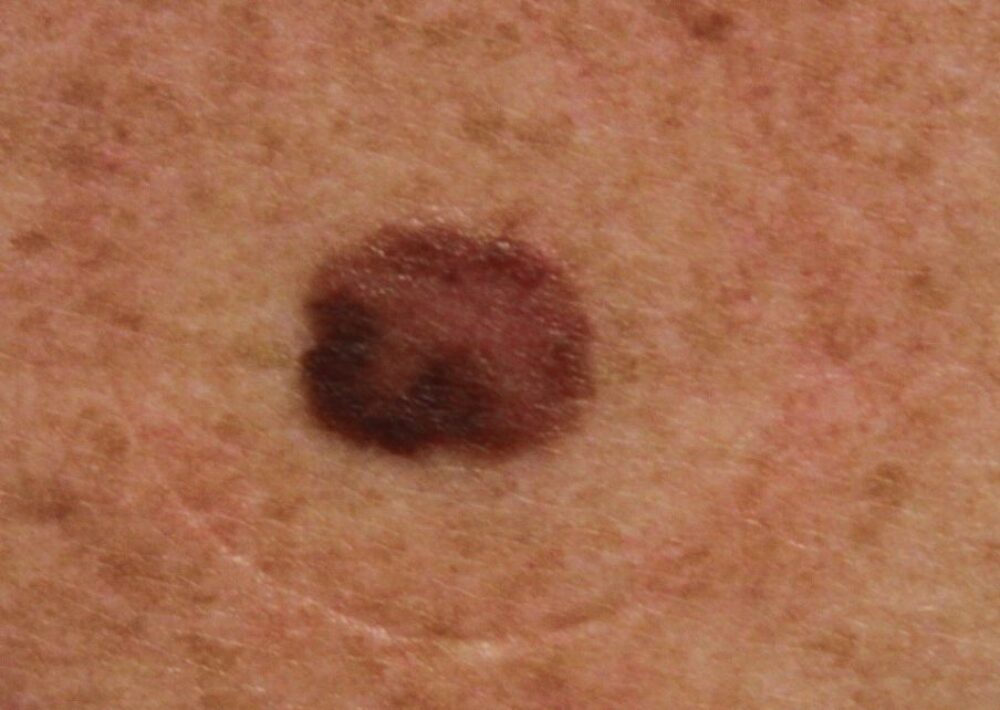
<p>This lady was referred with an asymptomatic lesion on the upper back. Given the site it was not possible to know how long the lesion had been present and whether it had changed. Clinically the lesion was an oval pink-brown macule, the colours were NOT well-organised. What is the differential diagnosis?</p>
Dermoscopy - Introduction, Terminology and Structures
A document to help you understand the dermoscopic descriptions used in this blog.
Dermoscopy Algorithm
To help you work through dermoscopic cases.
<p><strong>Clinically the lesion is suspicious as the two colours,</strong> pink and brown, are <strong>disorganised,</strong> as such the main differential in this case is between a dysplastic naevus and a melanoma.</p>
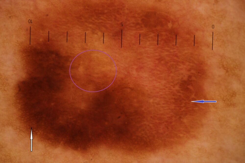
<p>Some mildly dysplastic naevi have two separate structures, and in some such lesions both structures take on a normal appearance within their own right, ie there is symmetry within asymmetry. With this lesion there is a normal looking brown network (white arrow), and a rather homogenous looking white network (blue arrow), however, there is a third structureless component (purple circle). Given that there are three different features (brown network, white network, structureless) there was more asymmetry then one would expect in a mildly dysplastic naevus and so the lesion was excised.</p>
<p>Histology showed a severely dysplastic naevus and so the case was discussed at the skin cancer MDT where a wide local excision of up to 5mm of normal surrounding skin was recommended.</p>
<p>Images copyright of South Tees Foundation Trust. </p>
