
<p>A 75 year old man was referred with a slow growing skin lesion. He had a previous history of melanoma and was immunosuppressed. </p>
Dermoscopy - Introduction, Terminology and Structures
A document to help you understand the dermoscopic descriptions used in this blog.
Dermoscopy Algorithm
To help you work through dermoscopic cases.
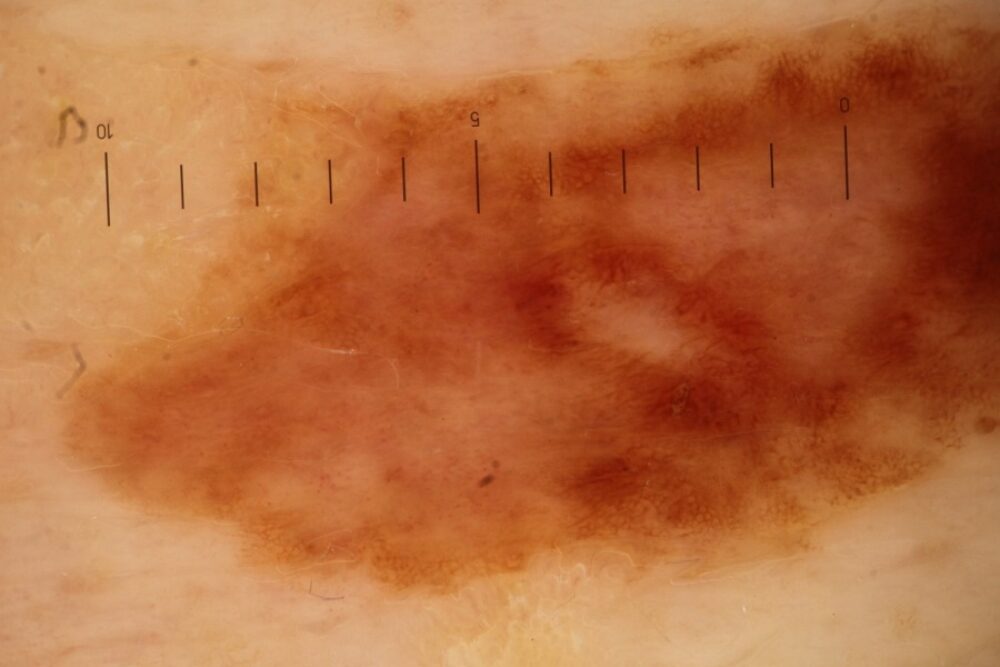
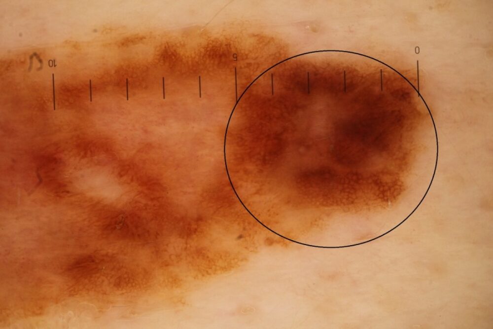
<p>Dermoscopically the lesion is asymmetrical with a brown pigment network and is clearly melanocytic. The lesion was excised, histology showed a melanoma. </p>
<p>Histology also showed areas of regression, but these features are not dermoscopically obvious. While there are several lightly coloured structureless areas these are no lighter than the surrounding skin, and similar changes can be seen in benign naturally regressing melanocytic naevi. Regression in melanoma is usually indicated by scar-like structureless areas that are paler than the surrounding skin, which may be associated with blue/grey structures and sometimes erythema. </p>
<p>Given the history of a changing melanocytic lesion in a 75 year old immunocompromised patient with a previous history of melanoma the decision to excise the lesion was taken before I examined dermoscopically. From a dermoscopic perspective the lesion is asymmetrical the right hand pole is certainly atypical. </p>
<p>Images copied with kind permission from South Tees Hospitals NHS Foundation Trust.</p>
<p>This will my last blog for 3 weeks as I am going on annual leave, in the meantime enjoy my blog on the <a href="http://www.aworldonthebrink.com/blog/the-farne-islands"><strong>Farne Islands</strong></a>. When I get back I will be writing about my experiences in the Canadian Rockies and Glacier National Park. </p>
