A 33 year old lady on melanoma follow-up was noted to have a changing lesion. What dermoscopic features can you see and what is the differential?
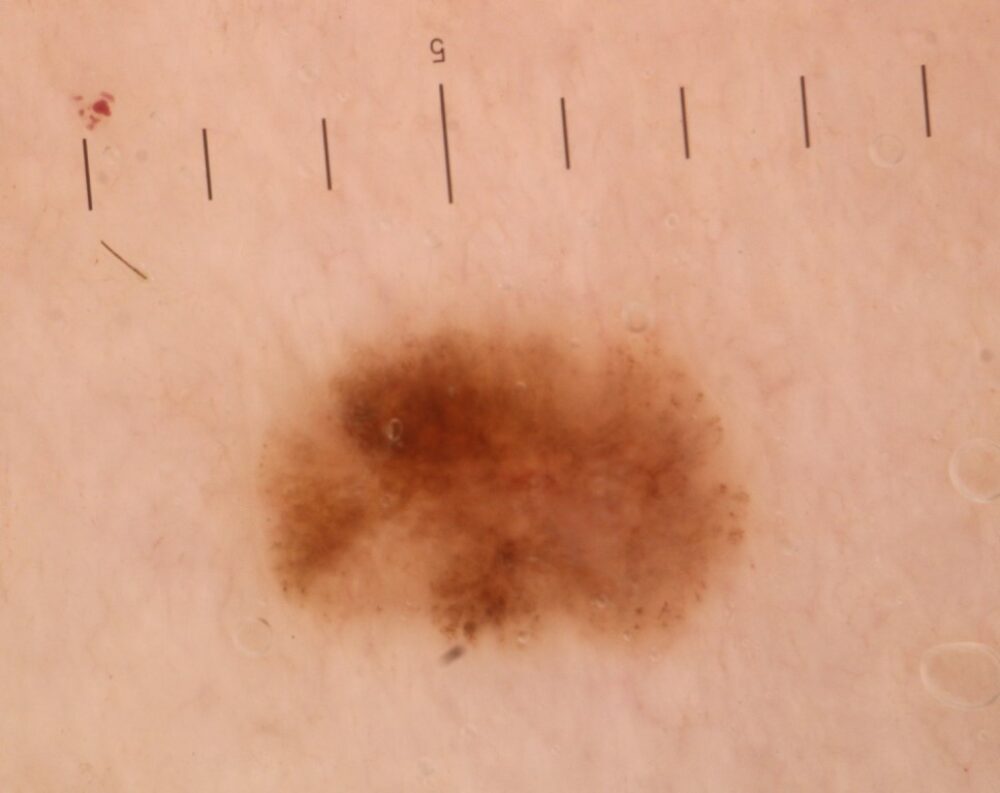
Dermoscopically there are grouped clods meaning the lesion is melanocytic. Using the associated algorithm there is asymmetry PLUS eccentric structureless areas (arrows), which on its own is of concern. In addition there is some variation in the clods of size, colour, and distribution (circles) in a patient with a past history of melanoma.
The lesion was excised with a histological diagnosis of melanoma in situ.

Dermoscopy - Introduction, Terminology and Structures
A document to help you understand the dermoscopic descriptions used in this blog.
Dermoscopy Algorithm
To help you work through dermoscopic cases.
Images copied with kind permission from South Tees Hospitals NHS Foundation Trust.