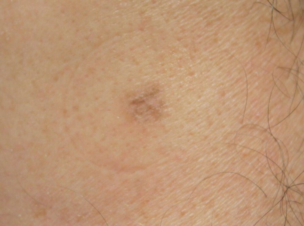
Looking at the clinical and dermoscopic images of this lesion on the right forearm what do you think the diagnosis is?

Dermoscopy - Introduction, Terminology and Structures
A document to help you understand the dermoscopic descriptions used in this blog.
Dermoscopy Algorithm
To help you work through dermoscopic cases.
Several lesions can have grey structures - lichenoid keratosis, basal cell carcinoma and lentigo maligna/melanoma.
This particular lesion can also be grey, and this is where the history is so important. This gentleman was referred with a slow growing raised lesion on the arm, from which the top had dropped off since being referred .... the diagnosis is a seborrhoeic keratosis, which commonly leaves behind grey structures where bits have come away.
Images copyright of South Tees Hospitals NHS Foundation Trust.