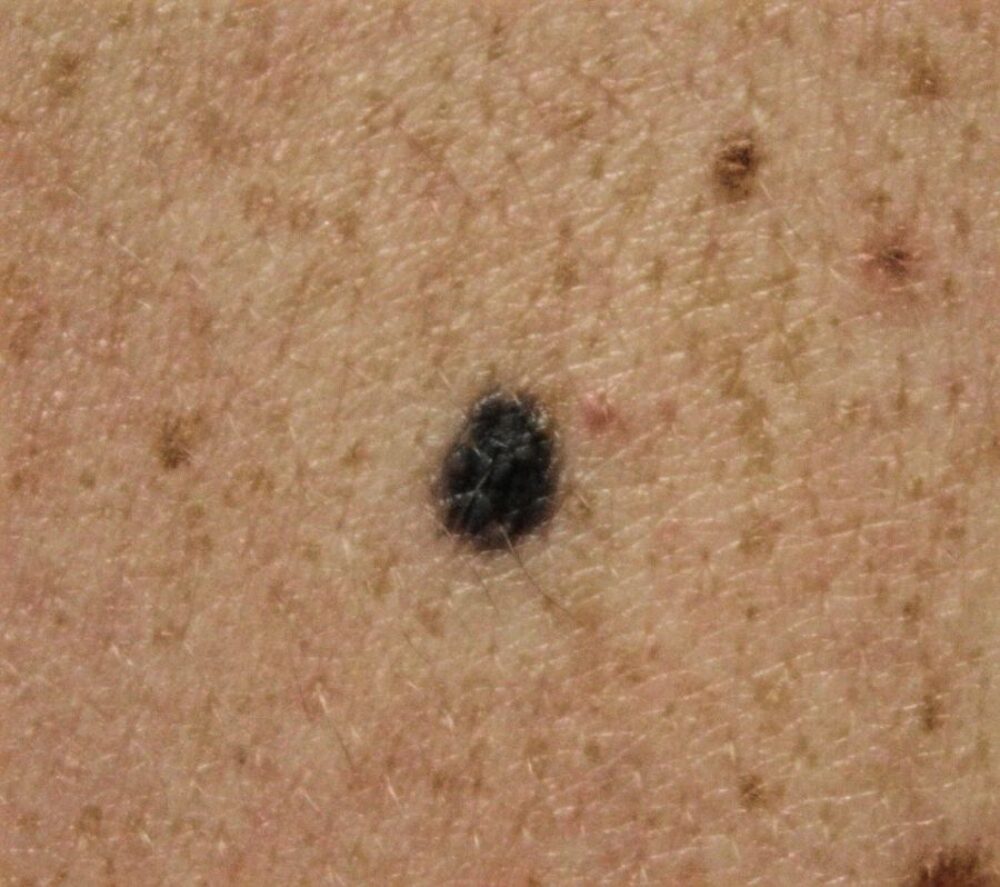
<p>This 63 year old man was referred with a lesion on his back, given the site he did not know whether it was new or long-standing. <strong>What colour do you see?</strong></p>
Dermoscopy - Introduction, Terminology and Structures
A document to help you understand the dermoscopic descriptions used in this blog.
Dermoscopy Algorithm
To help you work through dermoscopic cases.
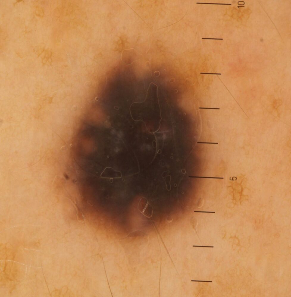
<p>This is a slightly asymmetrical lesion composed predominantly of a structureless area. Again <strong>what colour do you see?</strong><br></p>
<p>For me to be satisfied this was a blue naevus I would need to see blue or blue-grey, sometimes with a little white. I would also want to know that the lesion was not changing. The problem here was that we had no history of the lesion, and to me the colour looked more black with a little brown at the periphery, as such I could not exclude a melanoma in situ and so the lesion was excised.</p>
<p>The diagnosis was that of a blue naevus. </p>
<p>Blue naevi usually conform to the correct colour and history, but not always and occasionally lesions need excising. <strong>If we try and be too 'clever' with dermoscopy and only cut out the lesions that we are certain are cancerous then cases of skin cancer will be missed.</strong> </p>
<p>Copied with kind permission from South Tees NHS Foundation Trust. <br></p>
