
<p>A 57 year old patient was referred urgently with a changing pigmented lesion. Although there was no other skin history the patient was on long-term azathioprine. <strong>What is the most likely diagnosis?</strong></p>

<p>Dermoscopically there is asymmetry with an eccentric dark brown structureless area (white circle). There were also irregular brown-black clods (purple arrows) and a peripheral line (blue arrow). The lesion was excised, histology showed a <strong>melanoma in situ.</strong></p>
<p></p>
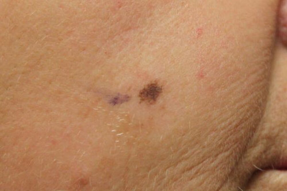
<p>Further examination also revealed an irregular <strong>brown patch on the face.</strong></p>
Dermoscopy - Introduction, Terminology and Structures
A document to help you understand the dermoscopic descriptions used in this blog.
Dermoscopy Algorithm
To help you work through dermoscopic cases.
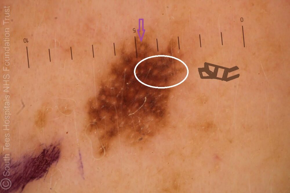
<p>On the face, brown-yellow circles are more reassuring than a <strong>brown or brown-black angulated network</strong>. In this case it was difficult to tell between the two patterns, however, if you inspect the area in the white circle (which I have projected to the right as a drawing ... I have never been the best at art), then you can see these structures are angulated. There was also a hint of grey in the upper pole. In addition the lesion is asymmetrical and the patient immunosuppressed, increasing the index of suspicion. Histology revealed a <strong>l</strong><strong>entigo maligna</strong>.</p>
<p>Always be aware that there may be more than one lesion of significance so a <strong>full skin examination</strong> is important. In terms of <strong>immunosuppression </strong>patients are at an increased risk of all types of skin cancer, although the risk is greatest for squamous cell carcinoma. </p>
<p>Images property of South Tees NHS Foundation Trust.<br></p>
