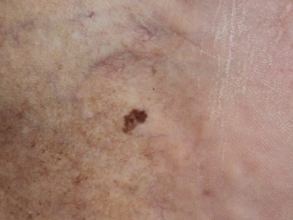
<p>This lesion on a heel was picked up by chance. The patient was unsure about the history. Clinically there was a little scale making a seborrhoeic keratosis part of the differential, but what do you think about the dermoscopic appearance?</p>

Dermoscopy - Introduction, Terminology and Structures
A document to help you understand the dermoscopic descriptions used in this blog.
Dermoscopy Algorithm
To help you work through dermoscopic cases.
<p>We can see <strong>aggregated brown clods</strong>, so as per the attached algorithm we need to manage this as probably melanocytic. Given the asymmetry and uncertain history excision was required.</p>
<p>Histology showed a seborrhoeic keratosis.</p>
<p>Dermoscopy is not perfect, but on a different day this could have been a melanoma. There remains no doubt that this lesion needed excising rather than observation. </p>
<p>Images copied with kind permission from South Tees Hospitals NHS Foundation Trust.</p>
<p>PS Happy New Year to you all. I am on holiday for the next couple of weeks :-) so my next blog will not be until later this month. </p>
