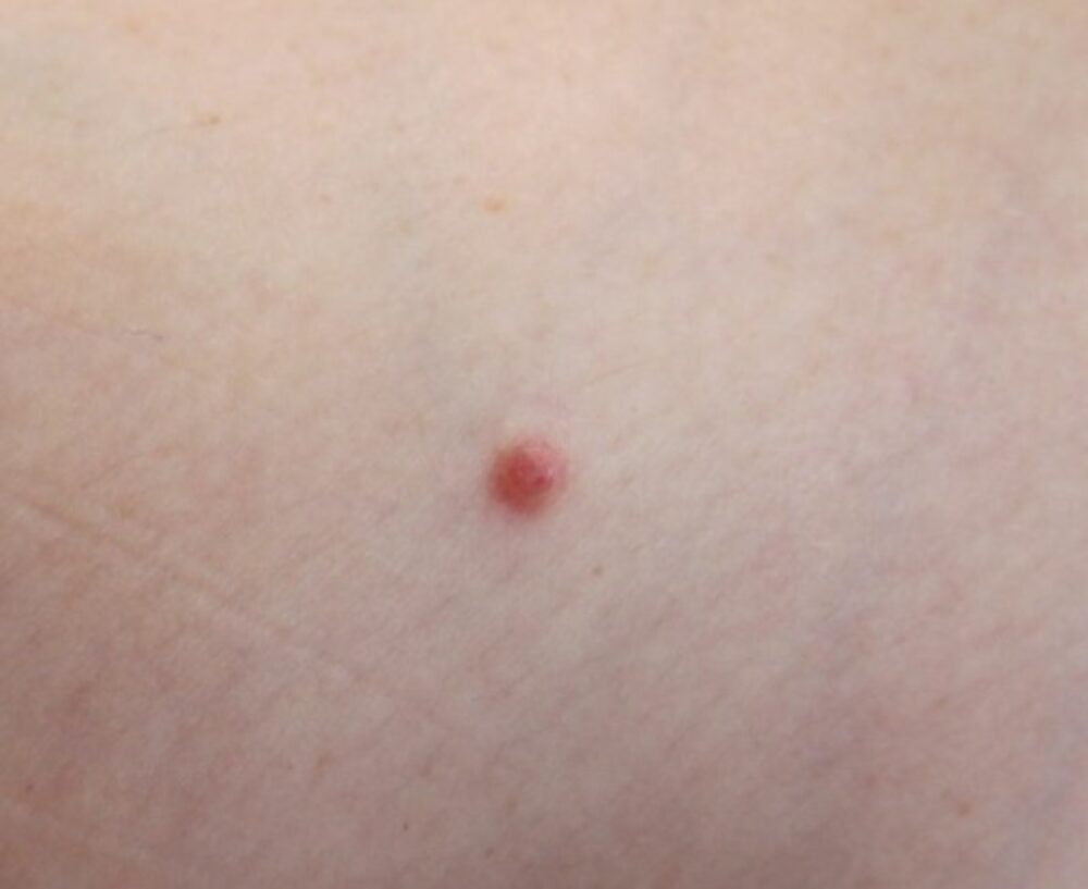
<p>A 31 year old lady presented with a new, asymptomatic, growing lesion under the elbow. There was no history of trauma and the lesion had not bled. What is the differential diagnosis? </p>
Dermoscopy - Introduction, Terminology and Structures
A document to help you understand the dermoscopic descriptions used in this blog.
Dermoscopy Algorithm
To help you work through dermoscopic cases.
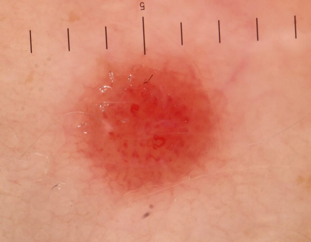
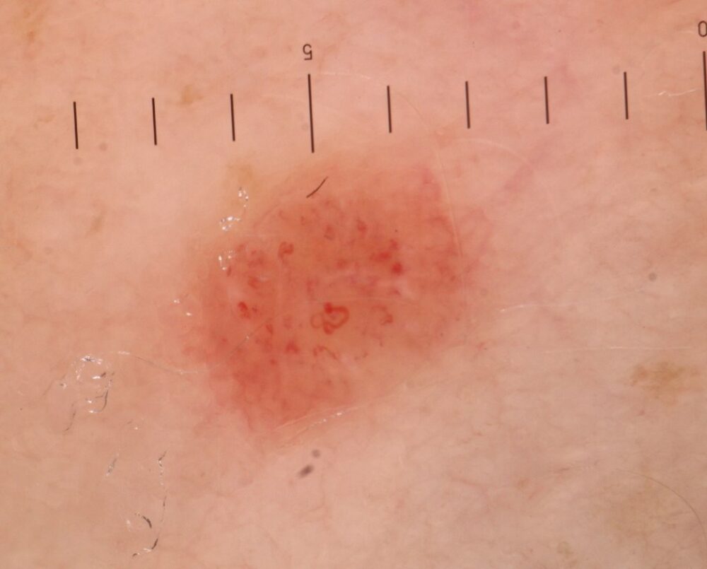
<p>When using contact dermoscopy the advice is to avoid exerting too much pressure otherwise the vessels are more difficult to make out. What is interesting here is that in the upper dermoscopic image using very limited pressure the vessels are harder to make out than compared with the lower dermoscopic image when a little extra pressure was exerted (however, if further pressure was applied I would have blanched the vessels all together). </p>
<p>Essentially there is a pink EFG (elevated, firm, growth - persistent) lesion that is dermoscopically structureless. These findings give cause for concern as the differential includes an amelanotic melanoma and a spitz naevus. Given the polymorphic nature of the vessels (ie significant variation in appearance) there was certainly concern about this being a melanoma.</p>
<p>Histology reported an atypical spitz naevus, which can be clinically and histologically difficult to differentiate from melanoma and so the lesion was treated with a respectable wide local excision. </p>
<p>Copied with kind permission from South Tees Hospitals NHS Foundation Trust. <br></p>
