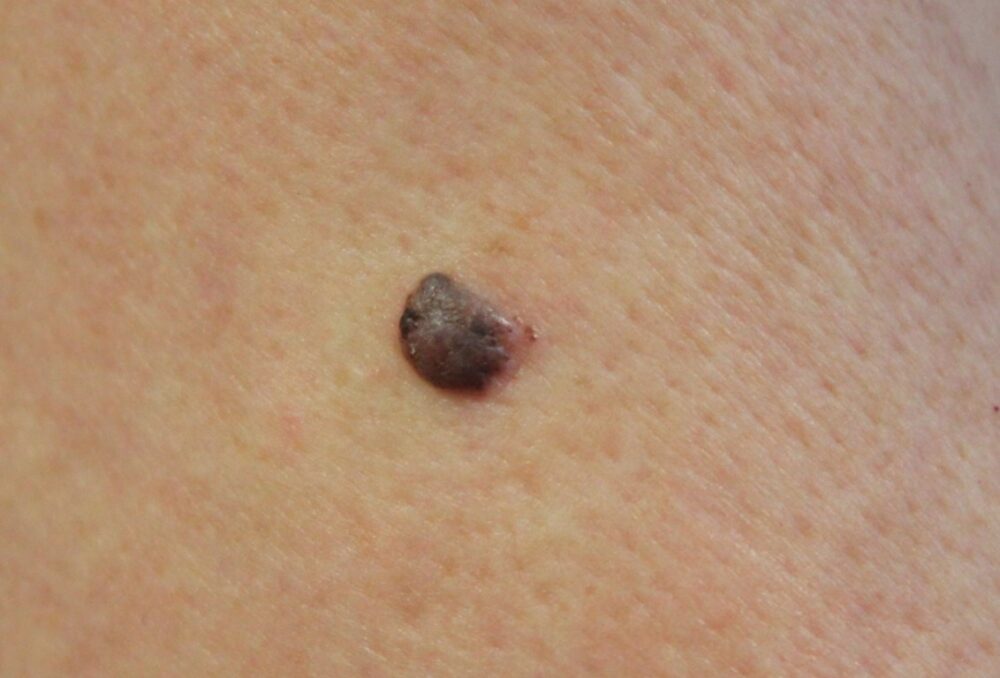
<p>What is the differential of this new solitary lesion on the arm?</p>
Dermoscopy - Introduction, Terminology and Structures
A document to help you understand the dermoscopic descriptions used in this blog.
Dermoscopy Algorithm
To help you work through dermoscopic cases.
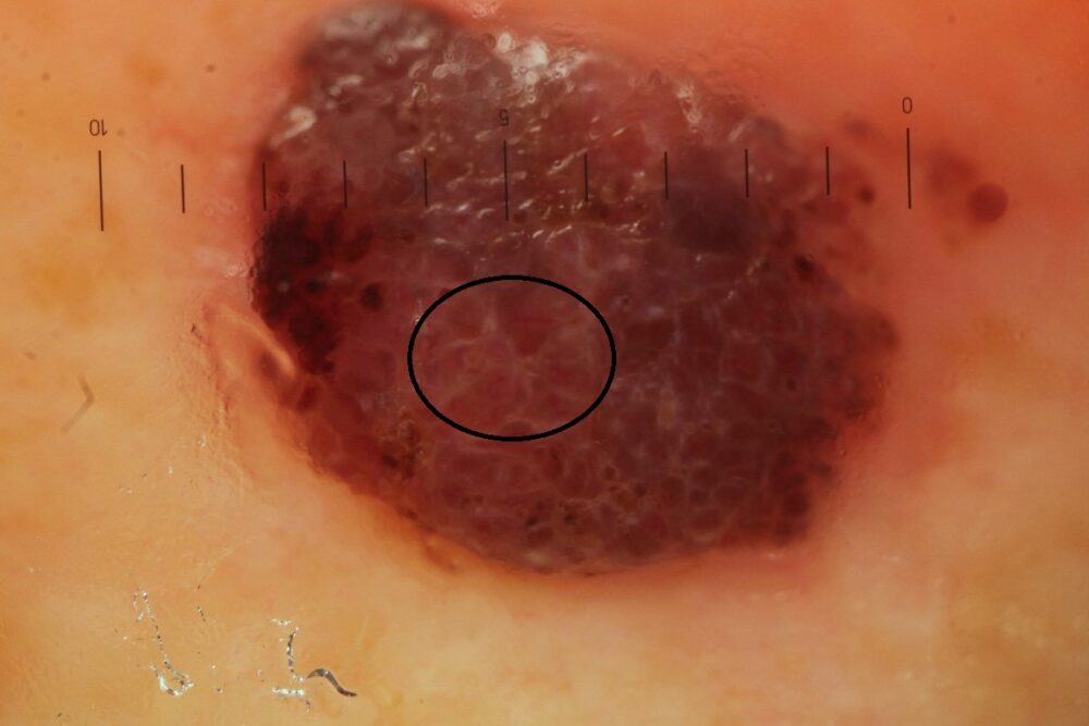
<p>Clinically the differential would have to include a seborrhoeic keratosis. Dermoscopically you could think about an angioma, but let us work our way logically through this case using the algorithm.</p>
<p>1) There are no obvious features that this is melanocytic</p>
<p>2) Are there any definitive features that this is benign? Clinically the differential would include a SK, and dermoscopically an angioma, however, put this together and things do not add up ie clinically this does not look like an angioma, and dermoscopically it does not look like a SK, and why has it suddenly grown? </p>
<p>3) There is no suggestion this is a BCC</p>
<p>4) Moving along to the end of the algorithm this leaves a relatively structureless lesion meaning that we can no longer exclude a melanocytic lesion. Given that this is a growing nodule there is no option other than to excise this immediately. <strong>Another concern is that a variety of vessels can be seen, some of which are linear, which should not be seen in angiomas </strong>(in the black circle ... not easy to see).</p>
<p>Histology showed a nodular melanoma, Breslow thickness 1.2 mm.</p>
<p><strong>The message here is that if we are faced with a structureless lesion that we can't diagnose clinically, we need to find out the diagnosis histologically.</strong> Some 50% of nodular melanomas are hypomelanotic. </p>
<p> Copied with kind permission from South Tees Foundation Trust.</p>
