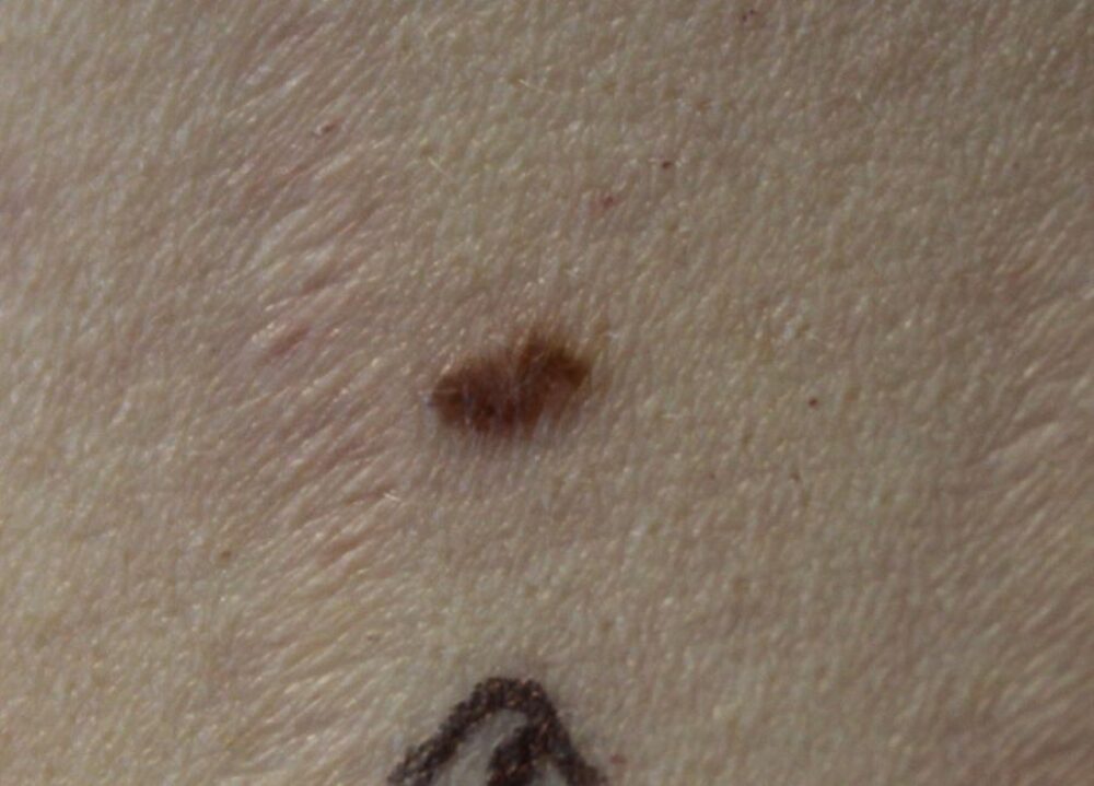
<p>A 27 year old lady was referred with a history of a changing lesion on her back. Clinically the lesion was a relatively symmetrical brown macule. What is the differential diagnosis?</p>
Dermoscopy - Introduction, Terminology and Structures
A document to help you understand the dermoscopic descriptions used in this blog.
Dermoscopy Algorithm
To help you work through dermoscopic cases.
<p>Clinically the lesion looked quite benign.</p>
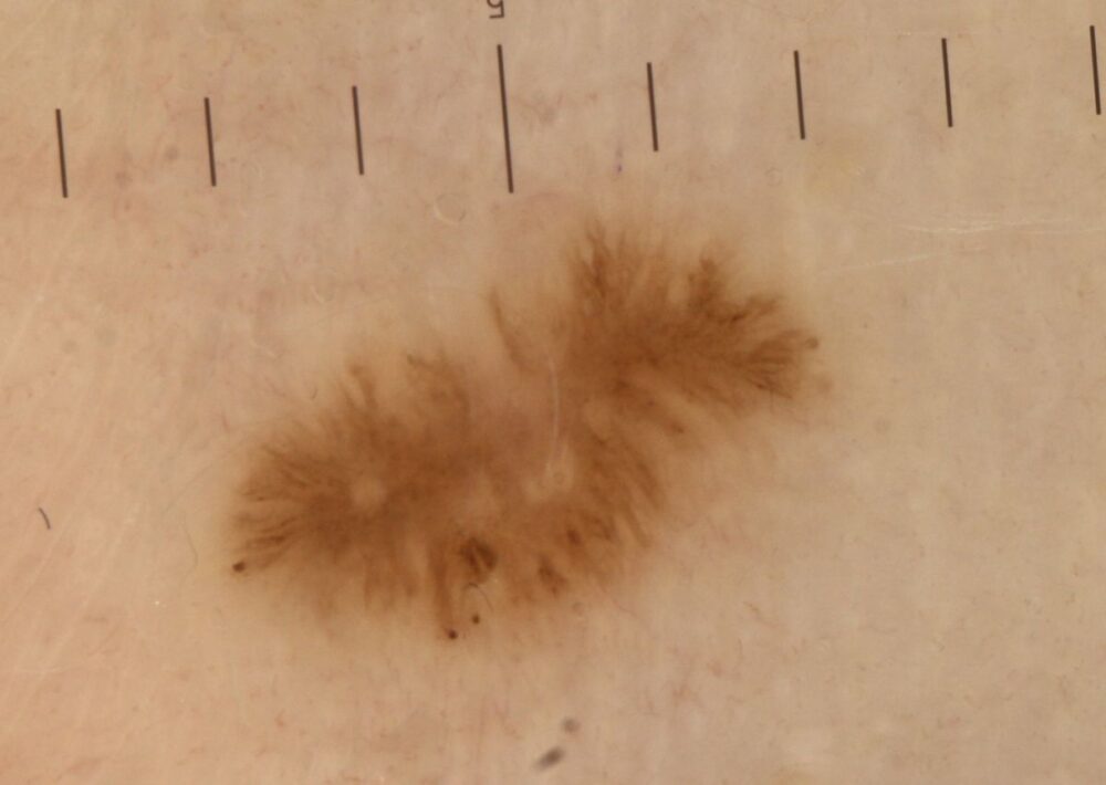
<p>Dermoscopically there are peripheral lines (streaks), which tells us that this is melanocytic. In addition the features are asymmetrical. As per the attached algorithm a combination of asymmetry and peripheral lines means that melanoma is possible. The differential also includes a pigmented spindle cell naevus of Reed, and given the clinical appearance a benign melanocytic naevus.</p>
<p>Histology revealed a benign melanocytic naevus. Retrospectively given the history and dermoscopic features the only safe option was to excise the lesion and send for urgent histology. I did not feel that monitoring was appropriate. Inevitably dermoscopy does not give all the answers and sometimes benign lesions need to be excised to be on the safe side. </p><p>Images property of South Tees NHS Foundation Trust.<br></p>
