Dermoscopy - Introduction, Terminology and Structures
A document to help you understand the dermoscopic descriptions used in this blog.
Dermoscopy Algorithm
To help you work through dermoscopic cases.
<p>Can you make out the diagnosis from the dermoscopic clues alone, and what do you think the history and clinical examination will be? <strong>(try not to scroll down to look at the clinical image first .... easier said than done!).</strong></p>
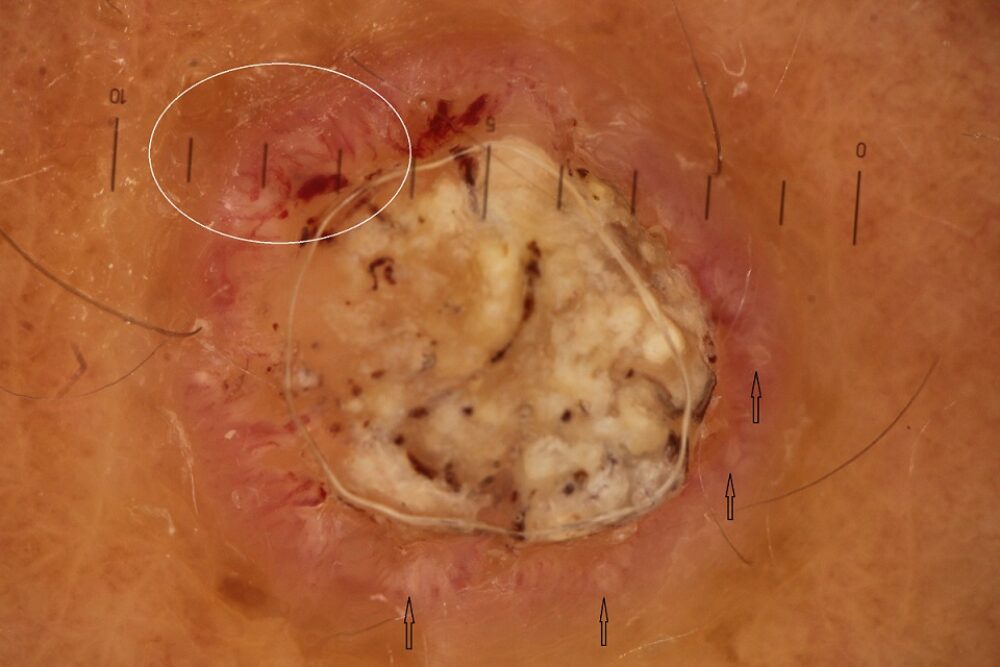
<p>Dermoscopically there is a large keratin core, suggesting an epidermal origin. There are also white-yellow peripheral clods (black arrows) and irregular vessels (white circle).</p>
<p>White/yellow clods can be found in a number of structures:</p>
<ul><li>Peripherally in Bowen's disease / SCC</li><li>Aggregated white clods in sebaceous gland hyperplasia</li><li>Scattered white clods (milia-like cysts) in seborrhoeic keratoses, BCC and rarely in melanocytic lesions</li></ul>
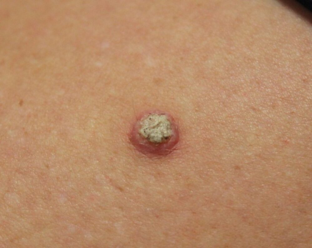
<p>There is too much central keratin for Bowen's so dermoscopically the diagnosis is consistent with the clinical image below of a well-differentiated SCC.</p>
<p>The lesion grew over 2-3 months, and was a little tender. Clinically there is a firm nodule with central scale. </p>