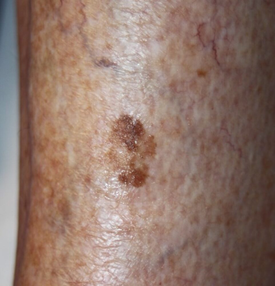
<p>A 76 year old woman was referred with a slow growing lesion on the shin. Clinically what is the differential?</p>
Dermoscopy - Introduction, Terminology and Structures
A document to help you understand the dermoscopic descriptions used in this blog.
Dermoscopy Algorithm
To help you work through dermoscopic cases.
<p>Clinically the lesion could be a solar lentigo/thin seborrhoeic keratosis or a superficial spreading melanoma.</p>
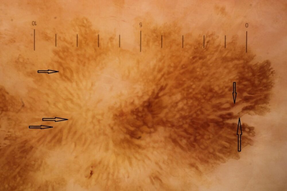
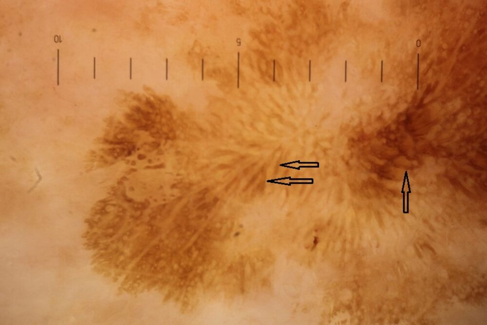
<p>Dermoscopically the colour is yellow-brown and well-demarcated. The primary structure are pigment bands (black arrows), a feature I first described in 2014. <strong>Pigment bands are thicker than lines, multiple and often curved, although can be straight. Features of a melanocytic lesion should be absent.</strong> Pigment bands will usually be found in thin lesions such as some solar lentigines/thin seborrhoeic keratoses, as was the case here. As lesions thicken the pigment bands take on the typical 3D appearance of fissures and ridges that we see in thicker seborrhoeic keratoses. </p>
<p>Images copied with kind permission from South Tees Hospitals NHS Foundation Trust.<br></p>
