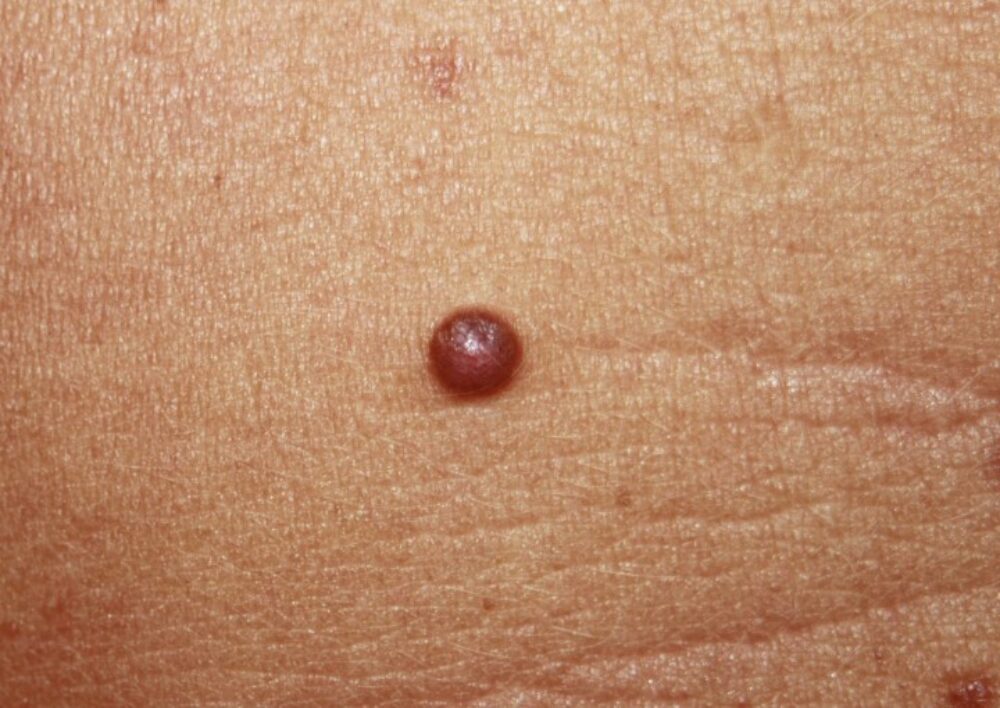
<p>This 27 year old lady presented with a lesion on the abdomen, there was no other relevant skin history. What is your initial clinical thinking, leave or excise? </p>

Dermoscopy - Introduction, Terminology and Structures
A document to help you understand the dermoscopic descriptions used in this blog.
Dermoscopy Algorithm
To help you work through dermoscopic cases.
<p>Clinically the lesion is highly symmetrical, but rather than being fairly soft, this was an <strong>EFG ... Elevated + Firm + Growth (sustained)</strong>, which should be a red flag. </p>
<p>Dermoscopcially there are aggregated clods in keeping with a melanocytic lesion. The clods are numerous and have with some variation in shape. The most striking feature is the presence of scattered short white lines that are reasonably asymmetrical. The few times I have seen this combination has resulted in the diagnosis of an atypical Spitz naevus, histology in this case was no different.</p>
<p>Such cases require MDT discussion, and while they are not categorised as skin cancer, their <strong>true malignant potential/biological behaviour is unknown</strong> with management usually recommending a wide local excision and a period of follow-up in line with melanoma. </p>
<p>Images copied with kind permission from South Tees Hospitals NHS Foundation Trust.</p>
