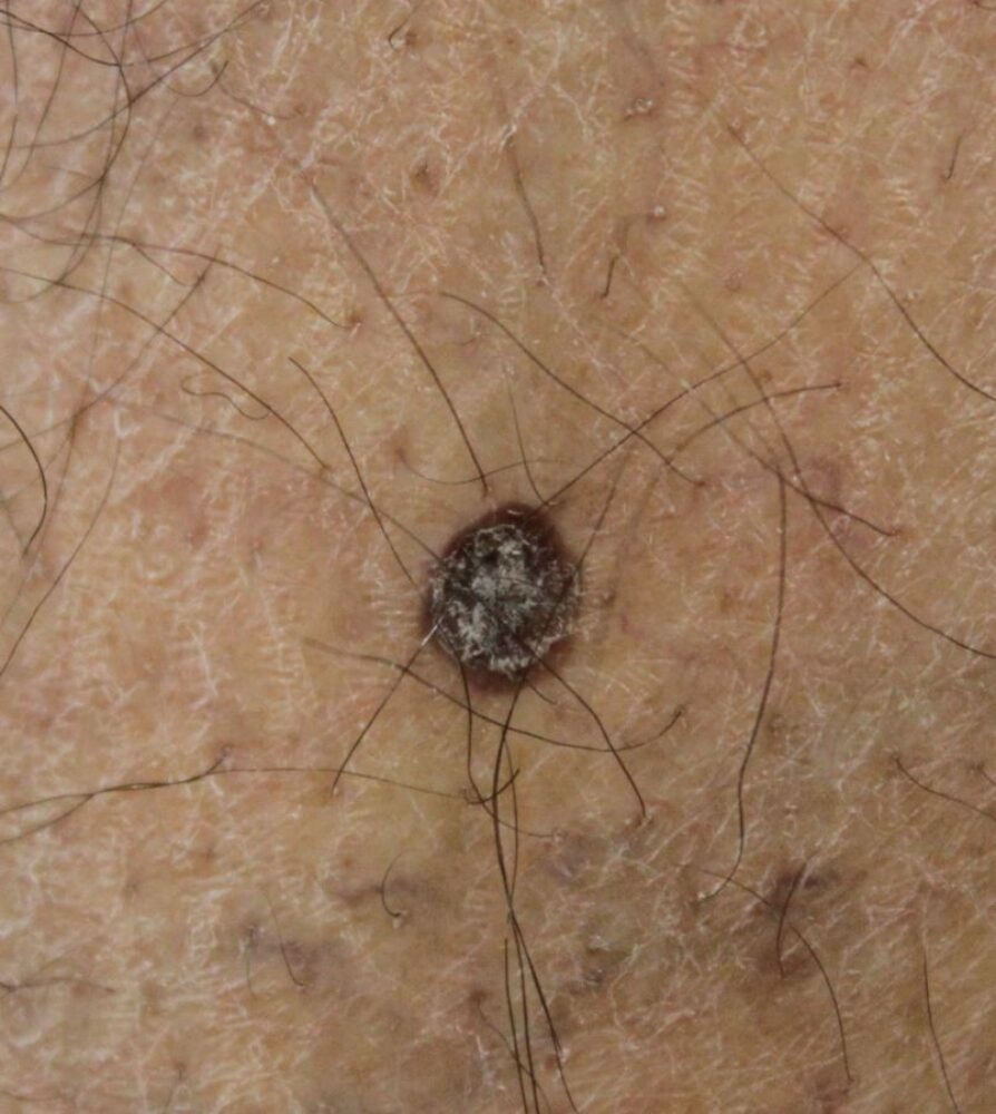
<p>A 67 year old patient with skin type V was referred with a new lesion on the shin. What is the differential? </p>

Dermoscopy - Introduction, Terminology and Structures
A document to help you understand the dermoscopic descriptions used in this blog.
Dermoscopy Algorithm
To help you work through dermoscopic cases.
<p>Dermoscopically we see radial brown lines in a starburst-like pattern, which is normally associated with a pigmented spindle cell naevus of Reed. Such lesions are normally found in younger patients and clinically I felt this was more likely to be non-melanocytic, indeed with the surface scale and the well-defined edge I favoured a thin SK. The options were to photograph and review in 3 months or take the liberty of a 3 mm punch biopsy - although the usual practice is to excise all suspect 'pigmented lesions' with a 2 mm clear margin, on occasions when the odds are very much against malignancy then in a specialist clinic a punch biopsy may suffice. I choose to do a punch biopsy that confirmed a thin SK.</p>
<p>I have since seen an identical lesion in a patient with skin type V, this time I was able to reassure without any intervention.</p>
<p>Copied with kind permission from South Tees Foundation Trust.</p>
