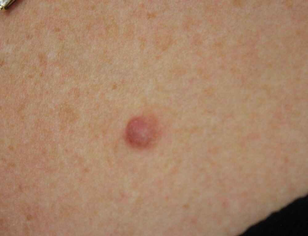
<p>A 46 year old lady was referred as a 2-week wait with a changing lesion on the left upper chest. The lesion had been present for some time, however, over the last 3 months it had appeared to grow. </p>
Dermoscopy - Introduction, Terminology and Structures
A document to help you understand the dermoscopic descriptions used in this blog.
Dermoscopy Algorithm
To help you work through dermoscopic cases.
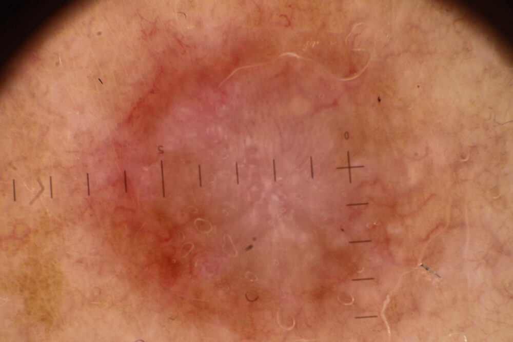
<p>Dermoscopy there are many white structures within the lesion, some of which are lines. White lines can be seen centrally in dermatofibromas, however, these were more extensive and asymmetrical then one would usually expect, and the lesion did not palpate like a dermatofibroma. Clinically this was an <strong>EFG (Elevated, Firm, Growing - persistent)</strong> lesion of uncertain aetiology, the rule for such lesions is that they should be referred as a 2-week wait and then excised for histology as a <strong>hypomelanotic melanoma is part of the differential</strong>. </p>
<p>Histology showed a benign appendageal tumour, namely a <strong>dermal duct tumour, </strong>which is a type of eccrine poroma located entirely in the dermis.</p>
<p>There are many different <a href="http://www.pcds.org.uk/clinical-guidance/appendage-tumours">appendageal tumours</a>, most of which are benign. The diagnosis of many appendageal tumours is usually only made by way of histology. </p>
<p>Copied with kind permission from South Tees Foundation Trust.</p>
