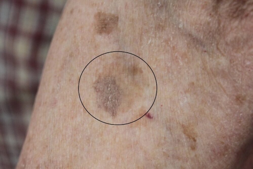
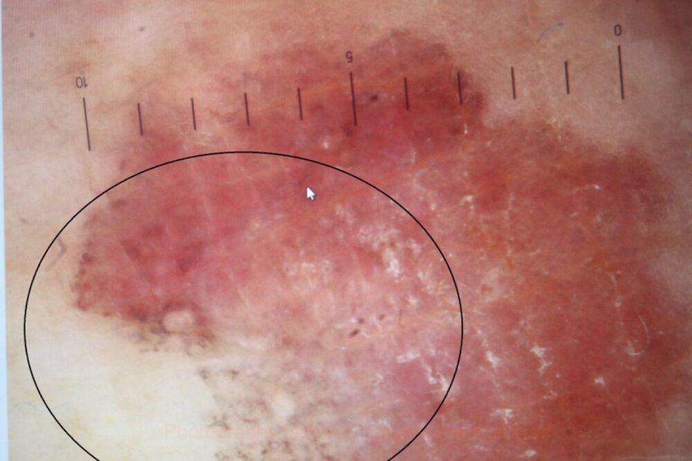
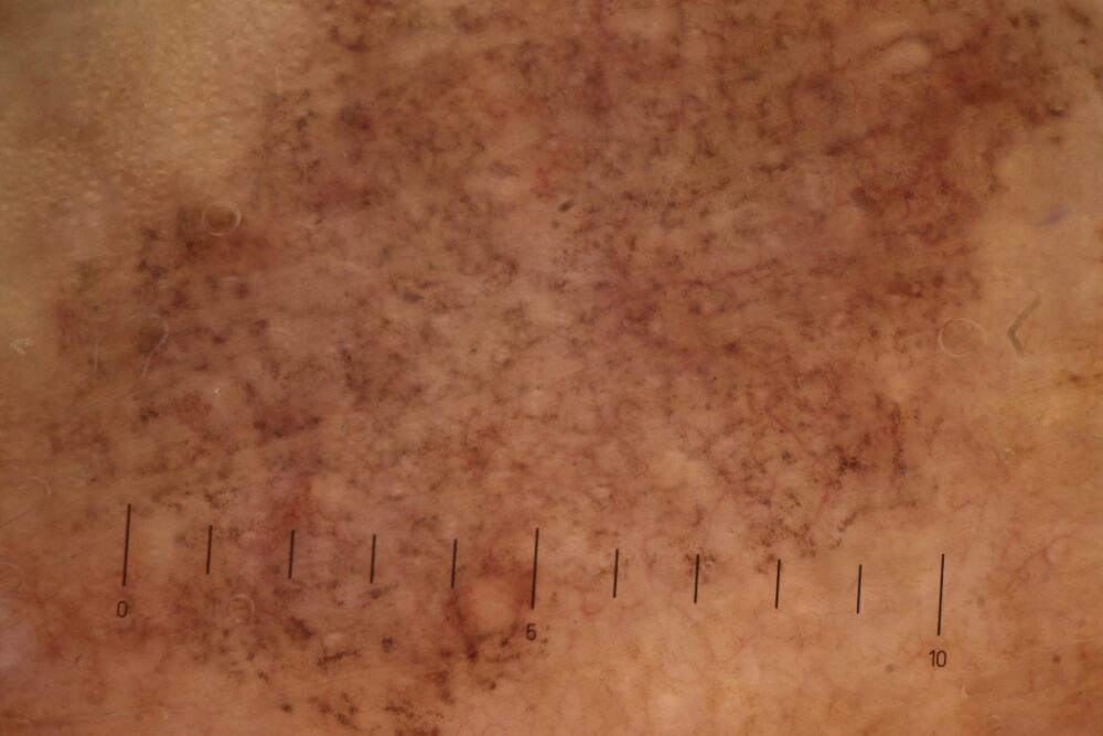
<p>A 73 year old presented with a long history of a flat brown lesion on the arm that became pink and a little itchy. The clinical image (left) is the end result, once the inflammation had settled. The first dermoscopic image below was the initial presentation, the second was taken a few weeks later at the same time that the clinical image was photographed.</p>
<p>The diagnosis is a lichenoid keratosis, which is a benign entity. </p>
<p>Lichenoid keratoses appear to result from the inflammatory destruction of a pre-existing epidermal lesion such as a solar lentigo or seborrhoeic keratosis. In the first dermoscopic image the lesion is clearly inflamed and a few grey dots are developing (circle). In the second image the inflammation has settled and there is a uniform distribution of grey dots.</p>
<p>Lichenoid keratoses are usually solitary and generally less than 1 cm in diameter. They are flat or slightly elevated. Lesions typically progress from an inflammatory phase, where there may be some pink-red discolouration, to a pigmented phase, where the lesion becomes grey-brown in colour and dermoscopically small grey dots can be seen distributed uniformly throughout the lesion.</p>
<p>For more information refer to the following link on <a href="http://www.pcds.org.uk/clinical-guidance/lichenoid-keratosis">lichenoid keratoses</a>. </p>
<p>Copied with kind permission from South Tees Hospitals NHS Foundation Trust. </p>
