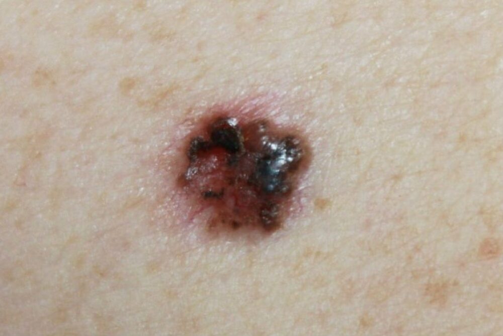
A 63-year old man presented with a lesion on his back of unknown history. On examination there was a shiny pigmented nodule. What is the diagnosis?
Dermoscopy - Introduction, Terminology and Structures
A document to help you understand the dermoscopic descriptions used in this blog.
Dermoscopy Algorithm
To help you work through dermoscopic cases.
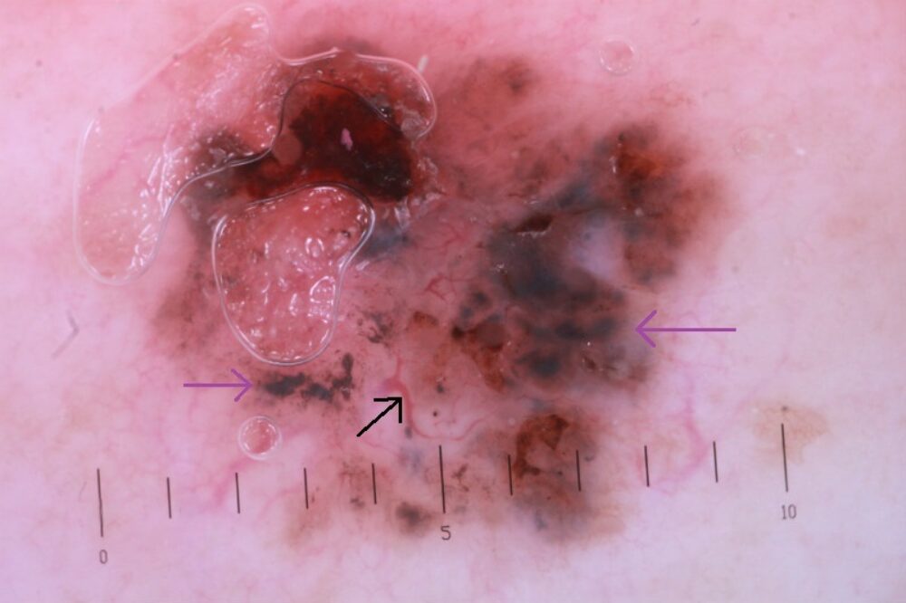
Clinically the differential included a basal cell carcinoma or a melanoma. Dermoscopically there is asymmetry with branching well-focused vessels (black arrow) and many blue clods (purple arrows) in keeping with a basal cell carcinoma - please refer to the algorithm. The lesion was excised with a 4 mm margin of surrounding skin.
Some BCC are heavily pigmented. If you have any doubt the patient should be referred as a 2-week wait.