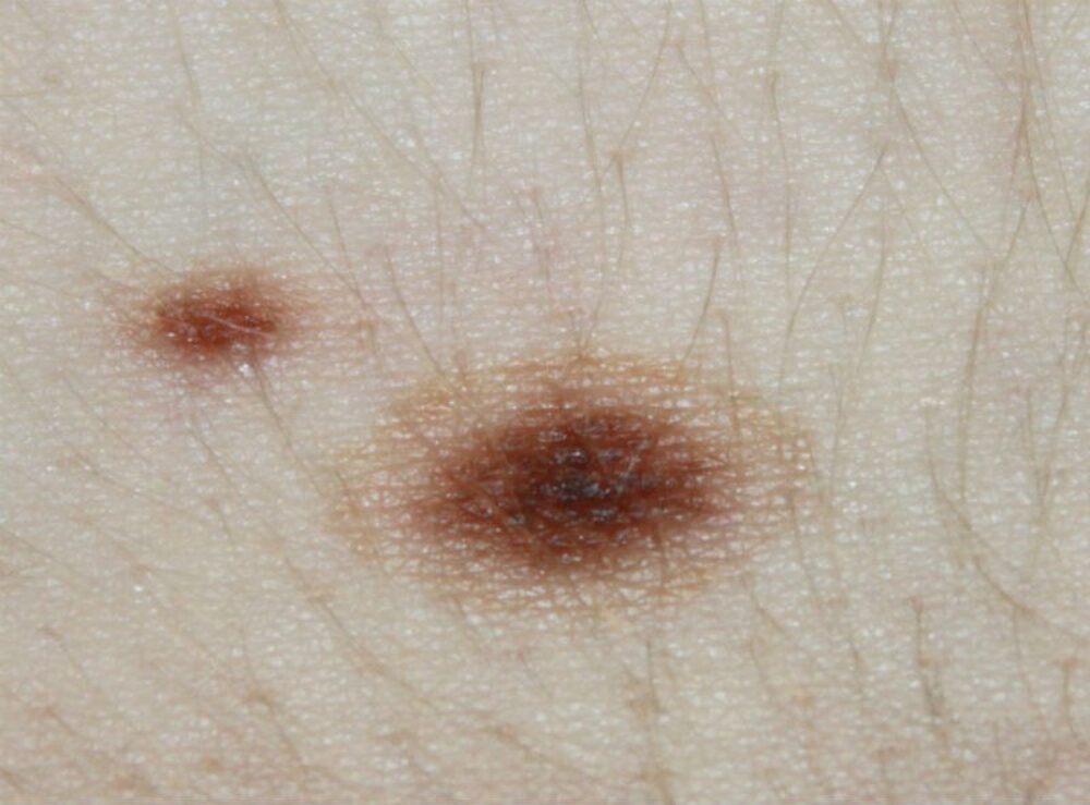
A 15 year old girl was referred with an enlarging lesion on the lower back. What is the diagnosis?

Dermoscopy - Introduction, Terminology and Structures
A document to help you understand the dermoscopic descriptions used in this blog.
Dermoscopy Algorithm
To help you work through dermoscopic cases.
Clinically there is a central brown macule surrounded by concentric circles of lighter and darker pigmentation giving the lesion a rosette-like appearance.
Dermoscopically the lesion has a central pigment network and is therefore melanocytic. There is a second network at the periphery (blue arrow). The features are highly symmetrical, which is reassuring. The diagnosis is a cockade naevus, a clinical variant of a benign melanocytic naevus. Such lesions usually arise in young patients, can be single or multiple, and most commonly involve the trunk although any site can be affected.
Images property of South Tees NHS Foundation Trust.