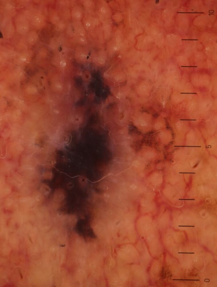
A 75 year old man was referred with a relatively new lesion on the left cheek. Based on the clinical and dermoscopic appearance what is the likely diagnosis?
Dermoscopy - Introduction, Terminology and Structures
A document to help you understand the dermoscopic descriptions used in this blog.
Dermoscopy Algorithm
To help you work through dermoscopic cases.
Clinically there is a small elongated black macule. Dermoscopically the lesion is mainly comprised of a black stuctureless area, and what is clearly apparent within this area is that there are no hair follicles, those that were present have been destroyed. Such findings are very suggestive of an invasive melanoma. Histology showed a superficial spreading melanoma with a relatively deep Breslow thickness of 2.2 mm (when compared with the small clinical size of the lesion).
Images copied with kind permission from South Tees Hospitals NHS Foundation Trust.