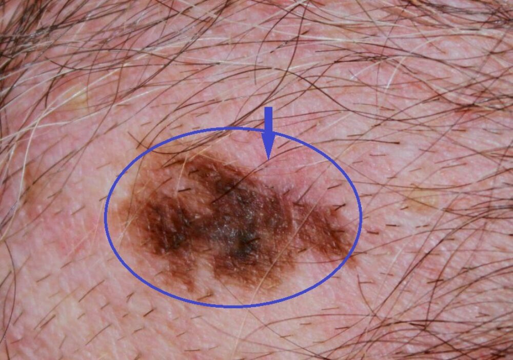
<p>A male patient was referred with what turned out to be a BCC on the left arm. He had worked outdoors and had extensive skin damage. A thorough skin examination revealed an irregular pigmented lesion on the chest wall. This had been present for many years with no obvious history of recent growth.</p>
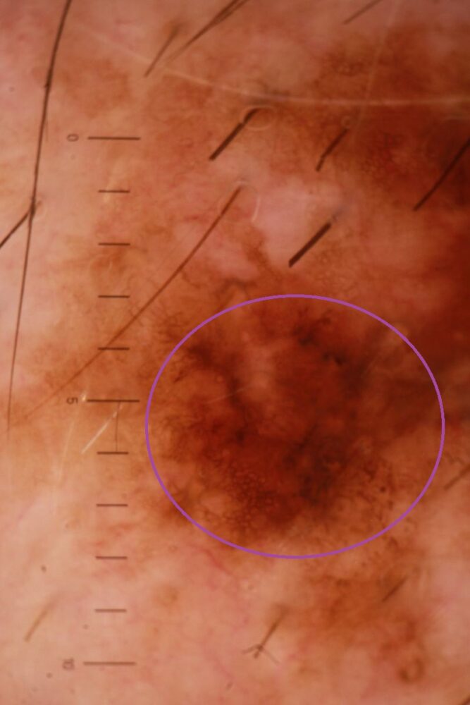
<p>Dermoscopy showed asymmetry and irregular grey structures (in the circle).</p>
Dermoscopy - Introduction, Terminology and Structures
A document to help you understand the dermoscopic descriptions used in this blog.
Dermoscopy Algorithm
To help you work through dermoscopic cases.
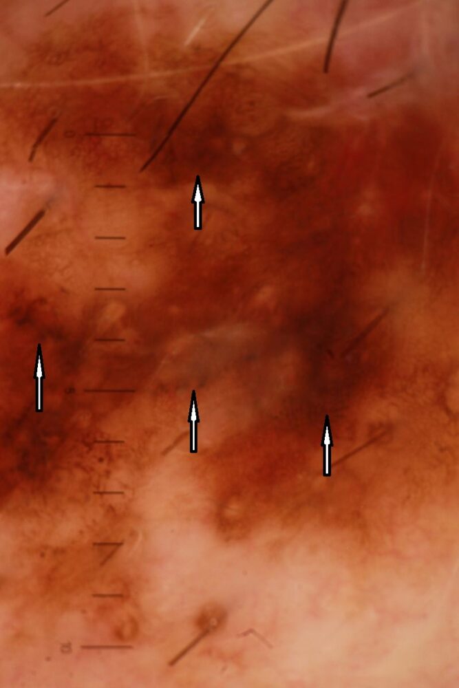
<p>More grey structures (arrows). As you will see from the algorithm, <strong>asymmetry plus grey structures is one of the red flags</strong>. Histology showed a <strong>lentigo maligna melanoma with regression</strong>.</p>
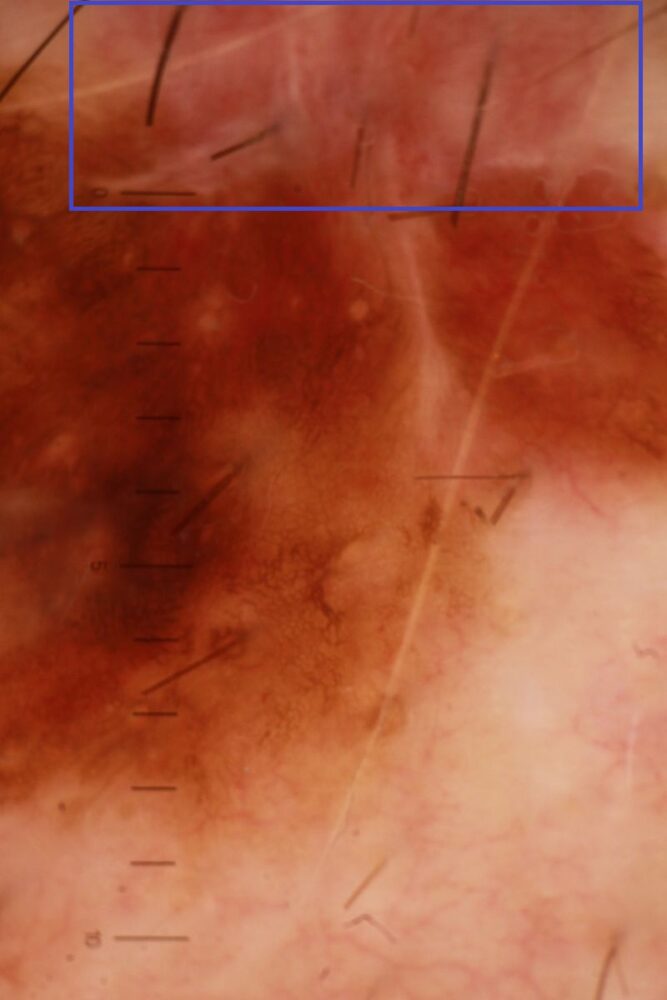
<p>It is likely that the <strong>area of regression</strong> is the area in the blue rectangle, which going back to the clinical image is the area highlighted by the blue arrow. The recommended wide excision was marked as 1 cm from the edge of clinically regressing margin.</p>
<p>A normal looking naevus that is not growing is nearly always going to be benign. However, <strong>be aware of the suspicious looking lesion (this was asymmetrical with an irregular border and had variation in colour) with no obvious history of change</strong> - thin melanoma's can grow very slowly, and those that are regressing may appear to be getting smaller. </p>
<p>Images copyright of South Tees Foundation Trust.</p>
