<p>A 72 year old lady was referred with a lesion on the bridge of the nose, which turned out to be an actinic keratosis. But what else did we find?</p>
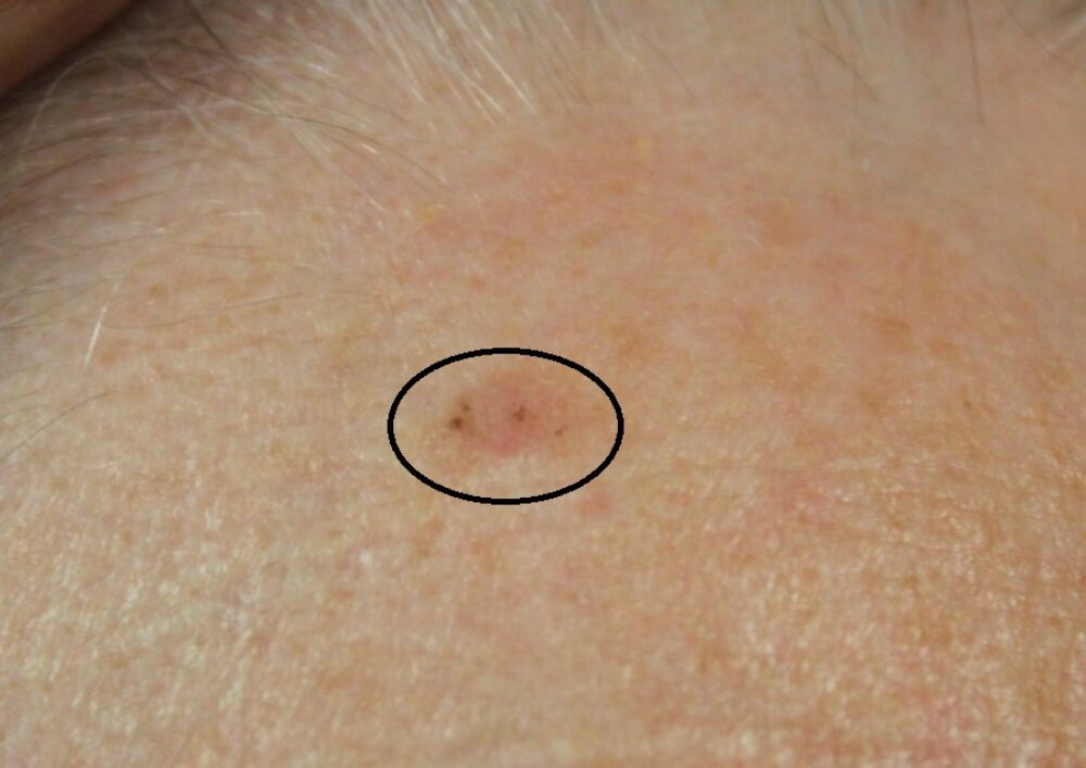
<p>There was an asymptomatic lesion on the right forehead. <strong>What is the differential?</strong></p>

<p>Clinically the lesion looked likely to be an actinic keratosis or a solar lentigo. However, dermoscopically there were <strong>peripheral brown irregular clods</strong> (purple arrows) and a <strong>small central ulcer</strong> (brown arrow) in keeping with a <strong>BCC</strong>, which in this case appeared relatively superficial. This dermoscopic appearance of some BCCs is one of the most difficult to recognise.</p>
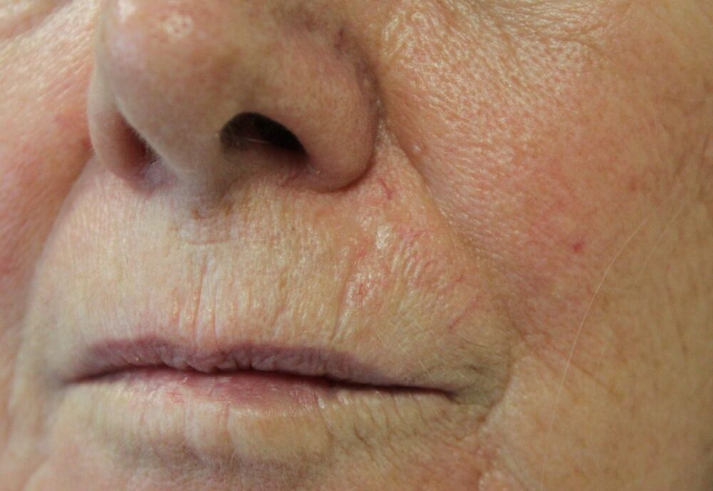
<p><strong>What else</strong> can you see above on the face?</p>
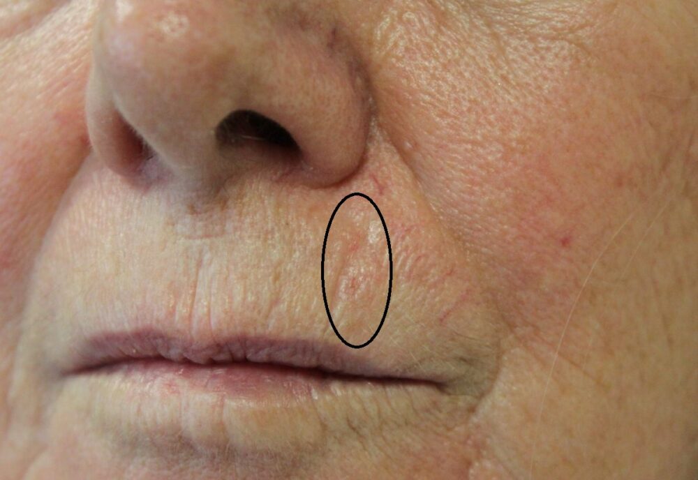
<p>There is an ill-defined <strong>shiny area</strong> of skin between the upper lip and nose. A shiny appearance may suggest a BCC, a good reason for having <strong>ample lighting</strong> when examining the skin. In poor light this would have been missed. The dermoscopic appearance follows.</p>

<p>Dermoscopy shows <strong>branching vessels </strong>(purple arrows), <strong>short white lines </strong>(black arrows) and a <strong>central erosion</strong> (brown arrow) in keeping with a BCC. The<strong> ill-defined </strong>edges seen clinically and dermoscopically point to this being <strong>infiltrative</strong>, such lesions can be a lot bigger than the naked-eye can see. Infiltrative BCC are most common on the central face, presenting as a slowly expanding white-yellow smooth patch/plaque, sometimes with no dermoscopic clues. Infiltrative lesions close to the eyes, nose or lips are best managed by <strong>MOHs surgery</strong>. </p>

<p>So what else could we find? A small pigmented lesion on the <strong>back</strong> under the bra-strap. <strong>What is the differential?</strong></p>

Dermoscopy - Introduction, Terminology and Structures
A document to help you understand the dermoscopic descriptions used in this blog.
Dermoscopy Algorithm
To help you work through dermoscopic cases.
<p>Clinically this could be melanocytic, a seborrhoeic keratosis or a BCC.</p>
<p>Dermoscopically there are <strong>fine branching (arborising) vessels</strong> (purple arrow) and <strong>blue clods</strong> (blue arrows) in keeping with a <strong>solid BCC</strong>.</p>
<p>It is always important to <strong>examine the facial skin using good light</strong>, and then examine <strong>the skin more widely</strong>. This patient had a superficial, solid, and infiltrative BCC, all of which may require different management. As you can see from the <strong>dermoscopic algorithm</strong> this patient had pretty much a full house of dermoscopic features found in BCC including fine branching vessels, blue clods, central erosions, irregular peripheral brown clods and short white lines.</p>
