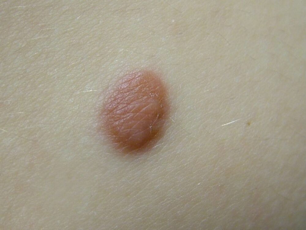
A 27 year old lady presented with a lesion on her left abdomen. This had been present for a number of years but she felt that it had recently got a little darker. What is the diagnosis?
Dermoscopy - Introduction, Terminology and Structures
A document to help you understand the dermoscopic descriptions used in this blog.
Dermoscopy Algorithm
To help you work through dermoscopic cases.
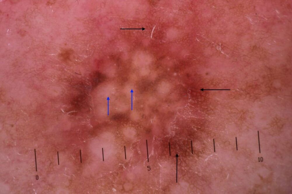
The predominant central features are white structureless areas (blue arrows), and the predominant peripheral feature is a brown rounded network (black arrows). The diagnosis is a dermatofibroma.
Dermatofibromas can often be diagnosed clinically as a firm smooth lump that can be palpated under the skin surface, lesions often dimple in the centre when squeezed.
Dermoscopically please refer to the algorithm. Most lesions have a central white component, unless they are flat in which case such a finding may be absent. The peripheral brown rounded network can be very subtle.