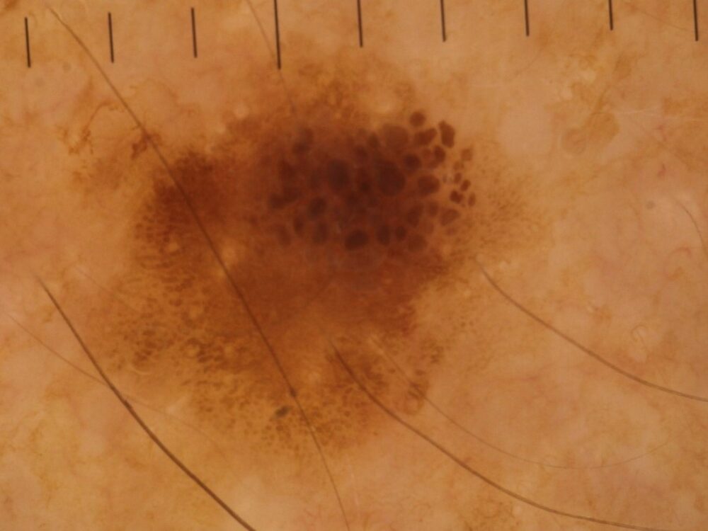
<p>Is this changing, slightly palpable lesion on the forearm of a 67 year old seborrhoeic, melanocytic or both?</p>
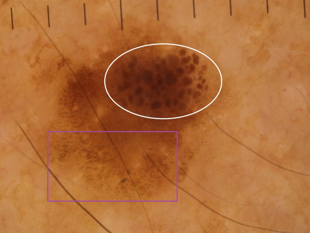
<p>The pigment bands (purple rectangle) are an evolving seborrhoeic keratosis, if left long enough these would become more elevated and appear as the fissures and ridges we see in some thicker SKs. <strong>The question is what are the brown clods (white circle)? </strong>In general, grouped smooth clods are seen in melanocytic lesions, and rough scattered clods in SK, so this should be a collision tumour with a melanocytic element .... which in this case could be of concern. Fortunately histology revealed this all to be seborrhoeic. </p>
<p>It is important to remember a dermatoscope is not a QR code reader, and that while dermoscopic structures and patterns usually follow what we have learnt, this is not always the case ..... the only thing that provided the definitive answer here was histology.</p>
