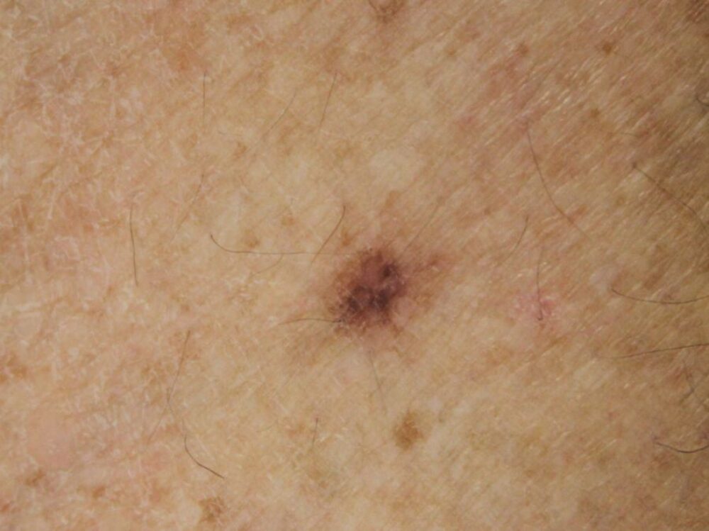
<p>A 72 year old lady was referred with a skin lesion on the shin that had possibly changed and become darker. What are the clinical differentials?</p>

Dermoscopy - Introduction, Terminology and Structures
A document to help you understand the dermoscopic descriptions used in this blog.
Dermoscopy Algorithm
To help you work through dermoscopic cases.
<p>There is a rounded network and a small hypopigmented area. The differential is between a melanocytic lesion and a dermatofibroma. Clinically the lesion was relatively flat with a regular firm lump that was palpated under the skin surface consistent with a dermatofibroma. <strong>Flatter dermatofibromas have a more substantial network (always rounded) with a structureless area that may be minimal or absent</strong>. A biopsy was not needed.</p><p>Copied with kind permission of South Tees NHS Foundation Trust.</p>
