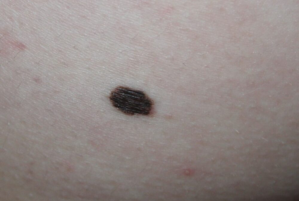
A 12 year old boy was referred with a clear history of a changing lesion on the left buttock. He was otherwise well. There was no relevant family history.
Dermoscopy - Introduction, Terminology and Structures
A document to help you understand the dermoscopic descriptions used in this blog.
Dermoscopy Algorithm
To help you work through dermoscopic cases.
How should this patient be managed?
Clinically the lesion is suspicious:
History of change
A = asymmetry
B = border, irregular
C = colour, different to his other naevi
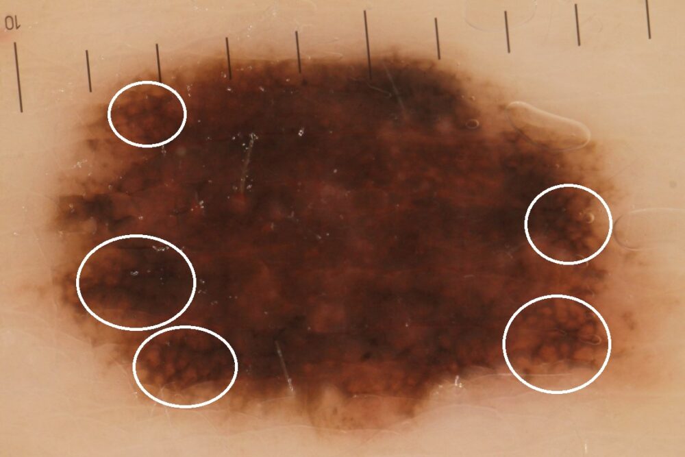
Dermoscopically there is also concern with asymmetry PLUS areas of an atypical black thickened network (white circles).
The lesion was excised. Histology revealed this was a benign melanocytic naevus, however, given the atypical clinical-dermoscopic appearance the sample was sent for a second opinion from which the conclusion was the same.
In reality melanoma under the age of 18 is rare, however, irrespective of age if a lesion looks suspicious the patient should still be referred as a 2-week wait, and as was the case here histology is often required to make certain of the diagnosis.
Copied with kind permission from South Tees Foundation Trust.