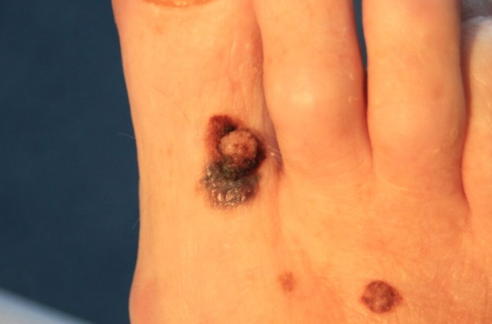
<p>This patient was referred with a suspicious lesion on the toe. A thorough examination revealed 3 other lesions that have also since been confirmed as melanoma. Let's have a look at the dermoscopic features.</p>
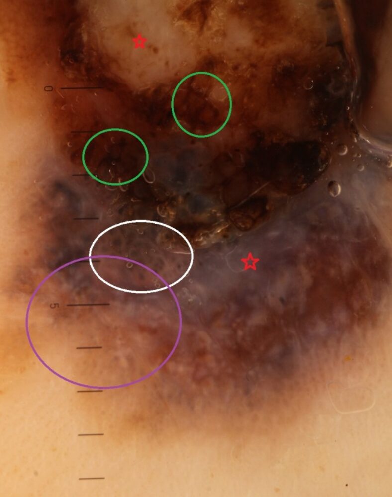
<p>- Asymmetry of structures </p>
<p>- Atypical network that is black, thickened, widely spaced (green circles)</p>
<p>- Irregular clods (white circle)</p>
<p>- Eccentric structureless areas. Upper area white, lower are blue-white suggesting possible regression (red stars)</p>
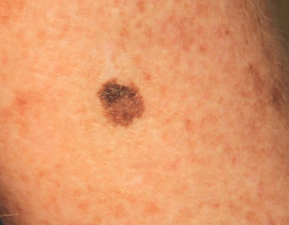
<p>This was the only one of the four that was in-situ.</p>
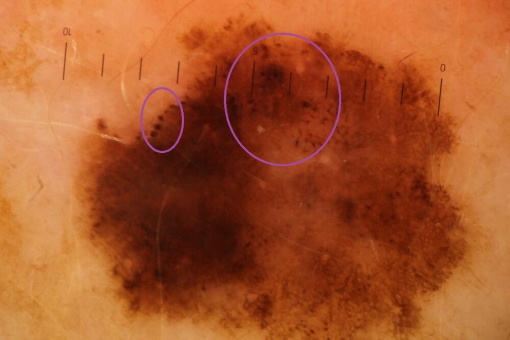
<p>- Multiple components with clods, network and structureless areas that are poorly organised resulting in marked asymmetry</p>
<p>- One of the main features is a very atypical pattern of clods with marked variation throughout the lesion in terms of colour (black clods highlighted in circles), size, shape and distribution <br></p>
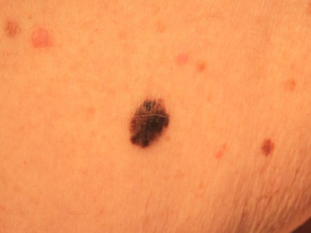
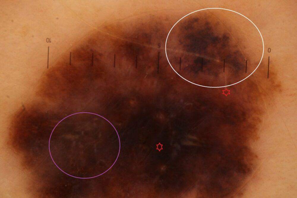
<p>- Asymmetry of structures</p>
<p>- Irregular grey-black structures (white circle)</p>
<p>- Irregular short white lines (purple circle). As discussed in previous blogs short white lines are not specific to melanocytic lesion, but there presence in such lesions is of concern </p>
<p>- Eccentric structureless areas (red stars)</p>
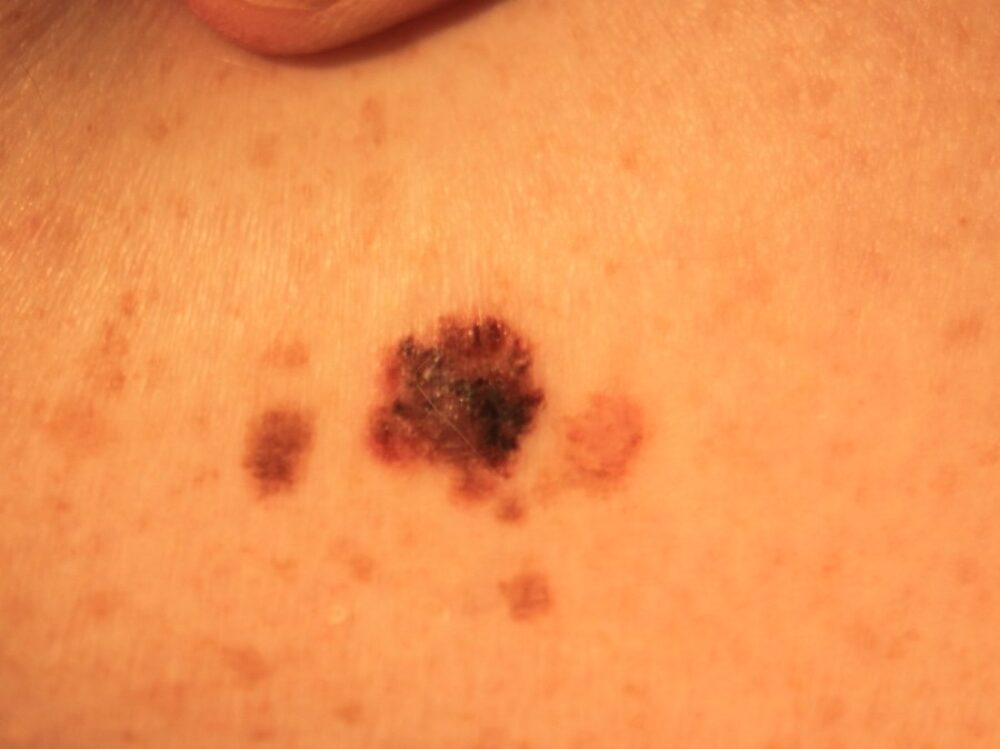

<p>- Asymmetry of structures</p>
<p>- Atypical pigment network (white circles)</p>
<p>- Eccentric multi-coloured structureless areas (purple circle)</p>
<p>- Irregular black clods (green circles) </p>
<p>Images copied with kind permission from South Tees Hospitals NHS Foundation Trust.</p>
