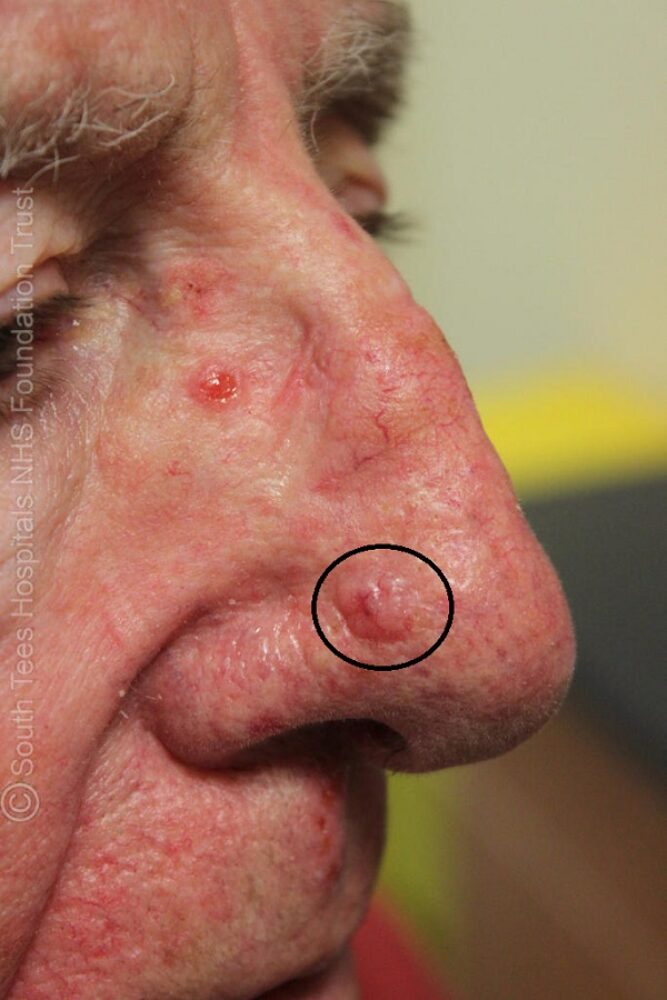
<p>This was the lesion that was referred. The easy one.</p>

<p>Dermoscopically many arborising vessels in keeping with a BCC.</p>

<p>The second lesion a little trickier.</p>

<p>Dermoscopically we see a central ulcer, but few other clues. What is the differential?</p>
<p>While a BCC is possible, a featureless ulcerated pink papule on the face could also be a moderate-poorly differentiated SCC. Histology revealed a BCC.</p>
<p>So we find BCC on the face, what do we always do next .... a general skin examination. </p>

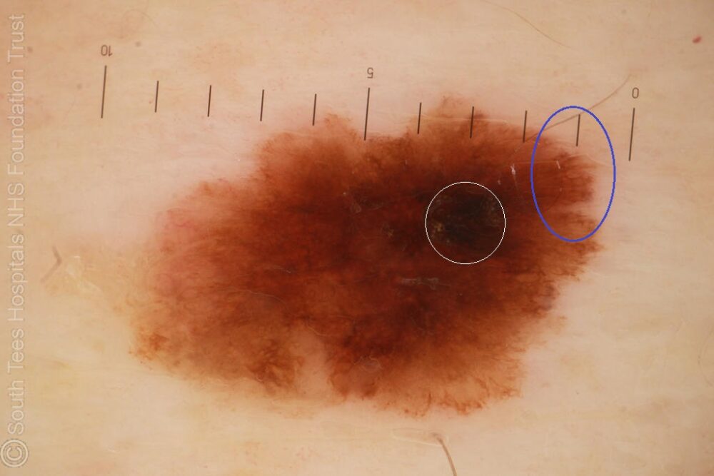
<p>The patient was unaware of this lesion on his abdomen. </p>
Dermoscopy - Introduction, Terminology and Structures
A document to help you understand the dermoscopic descriptions used in this blog.
Dermoscopy Algorithm
To help you work through dermoscopic cases.
<p>This can't be normal - marked asymmetry, eccentric black structureless area (white circle) and peripheral lines (blue circle). Histology showed a severely dysplastic naevus, however, this could easily have been a melanoma dermoscopically, and certainly if it had not been excised it would have become life-threatening.</p>
<p><strong>When commissioning skin cancer pathways make certain that as part of the contract specialists must undertake a thorough skin examination in patients found to have pre-cancerous lesions and/or skin cancer, you may be amazed how many services don't ...... such a policy saves lives.</strong></p>
<p>Images copied with kind permission from South Tees Hospitals NHS Foundation Trust.</p>
